Event submissions
Published
This submission belongs to the session S3. Acute Lymphoblastic Leukemia of the event The 1st International Online Conference on Hematology
Published date
23 Oct, 2026
Academic Editor
 Claudio Cerchione
Citation
Vira Anatoliyivna Barilka, Volodymyr Lvovych Matlan, Olha Oleksiyivna Shalay, Sofiya Vasylivna Prymak, Vasyl Leonidovych Novak, The Role of Transforming Growth Factor Beta 1 in Tumor Microenvironment of Acute Lymphoblastic Leukemia Patients, in Proceedings of The 1st International Online Conference on Hematology, 28 October–30 October 2026, MDPI: Basel, Switzerland
Share
Email
Facebook
Twitter
LinkedIn
The Role of Transforming Growth Factor Beta 1 in Tumor Microenvironment of Acute Lymphoblastic Leukemia Patients

Vira Anatoliyivna Barilka 1
Volodymyr Lvovych Matlan 2
Olha Oleksiyivna Shalay 1
Sofiya Vasylivna Prymak 3
Vasyl Leonidovych Novak 4
1. Laboratory of Immunology and Genetics of Blood Neoplasms, State Institution "Institute of Blood Pathology and Transfusion Medicine, National Academy of Medical Science of Ukraine, Lviv, 79044, Ukraine
2. Non-Profit State Company, "Danylo Halytsky Lviv National Medical University", Lviv, 79010, Ukraine
3. Department of Hematology, State Institution "Institute of Blood Pathology and Transfusion Medicine, National Academy of Medical Science of Ukraine, Lviv, 79044, Ukraine
4. State Institution "Institute of Blood Pathology and Transfusion Medicine, National Academy of Medical Science of Ukraine, Lviv, 79044, Ukraine
Abstract
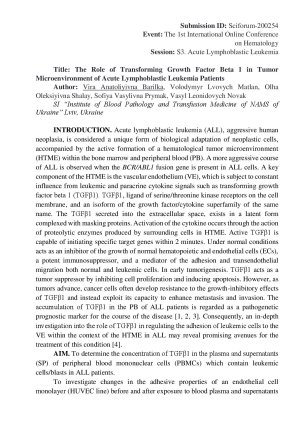
For this purpose: determined the concentration of TGFβ1 in plasma and supernatant (Sp) of mononuclear cells of peripheral blood (PBMC) of patients (pts) with acute lymphoblastic leukemia (ALL); and estimated the effect of these samples on the change the adhesiveness of endothelial cell, HUVECs, in vitro, by adhesion of K562 cells with positive expression of BCR/ABL1) and L1210 cells line with oncogene c- Myc; and the obtained results analyzed in relation to the parameters of the healthy persons. The median age was 56 years. The number of blasts in the PB - 55,4 ± 9,71 × 10 9/L, which was the basis for considering this PB as a tumor microenvironment of acute lymphoblastic leukemia (TME). Cells lines K562 and L1210 were labeled with a 3H-methylthymidine, and not treated with cytokines before the investigation. The concentration of TGFβ1 was determined by a biological method.
Results. The concentration of TGFβ1 in the plasma of pts was higher than in the control (p<0.001). An inverse correlation was established between the level of TGFβ1 in the plasma and the number of blasts in PB of pts (r = – 0.40). TGFβ1 in the Sp of pts was higher (p<0.05) than in the plasma of pts, according to this, PBMC were the source of TGFβ1 in ALL pts. The K562 cells demonstrated higher adhesion compared to L1210 after treatment of HUVEC with plasma and Sp of PBMC ALL pts (p < 0.05; in both cases). A negative correlation was found between the concentration of TGFβ1 in the plasma of pts and the adhesion of K562 (r = – 0.56) and L1210 (r = – 0.30) to HUVECs monolayer.
The results demonstrated the ability of HUVECs differentially form adhesive contacts, depending on the type of origin of the leukemic cells and TGFβ1 level in the TME of ALL.
Keywords
Acute Lymphoblastic Leukemia
TGFβ1
Adhesion
L1210
K562 Leukemic Cells Lines
HUVECs
TME
Poster
Barilka V. Poster Submission ID Sciforum-200254.pdf
Masked Acute Promyelocytic Leukemia with Monosomy 17 and Marker Chromosome: Importance of Multimodal Molecular Diagnosis