Event submissions
EventsThe 2nd International Electronic Conference on Medicinal Chemistry and Pharmaceutics
Published
This submission belongs to the session S4. Natural Products and Biopharmaceuticals of the event The 2nd International Electronic Conference on Medicinal Chemistry and Pharmaceutics
Published date
28 Oct, 2026
Academic Editor
 Alfredo Berzal-Herranz
Contribution&Funding
Conceptualization,
[Vagif A. Gasanov]
Methodology,
[Dmitry A. Kashirskikh]
Formal analysis,
[Victoria A. Khotina] ; [Arthur A. Lee]
Investigation,
[Arthur A. Lee] ; [Olesya O. Klychkova] ; [Margarita P. Markina] ; [Vitalia S. Novikova] ; [Victoria A. Khotina] ; [Dmitry A. Kashirskikh]
Data curation,
[Arthur A. Lee] ; [Olesya O. Klychkova] ; [Margarita P. Markina] ; [Vitalia S. Novikova] ; [Dmitry A. Kashirskikh]
Writing - original draft,
[Victoria A. Khotina]
Writing - review,
[editing] ; [Dmitry A. Kashirskikh] ; [Vagif A. Gasanov]
Visualization,
[Victoria A. Khotina]
Supervision,
[Vagif A. Gasanov]
Project administration,
[Vagif A. Gasanov]
Funding acquisition,
[Vagif A. Gasanov]
Funding: The Ministry of Science and Higher Education of the Russian Federation in 2026 (No. 0088-2025-0002).
Citation
Arthur A. Lee, Olesya O. Klychkova, Margarita P. Markina, Vitalia S. Novikova, Victoria A. Khotina, Dmitry A. Kashirskikh, Vagif A. Gasanov, DESIGN AND IN VITRO FUNCTIONAL EVALUATION OF M-6HIS-GLP-1-6HIS, A DUAL-HEXAHISTIDINE-TAGGED GLP-1 VARIANT, in Proceedings of The 2nd International Electronic Conference on Medicinal Chemistry and Pharmaceutics, 1 November–30 November 2026, MDPI: Basel, Switzerland
Share
Email
Facebook
Twitter
LinkedIn
DESIGN AND IN VITRO FUNCTIONAL EVALUATION OF M-6HIS-GLP-1-6HIS, A DUAL-HEXAHISTIDINE-TAGGED GLP-1 VARIANT

Arthur A. Lee 1
Olesya O. Klychkova 1
Margarita P. Markina 1
Vitalia S. Novikova 1

Victoria A. Khotina 1
Dmitry A. Kashirskikh 1
Vagif A. Gasanov 1
1. Core Centrum «Genomic Technologies Group», Koltzov Institute of Developmental Biology of the Russian Academy of Sciences (IDB RAS), Moscow, 119334, Russia
Abstract
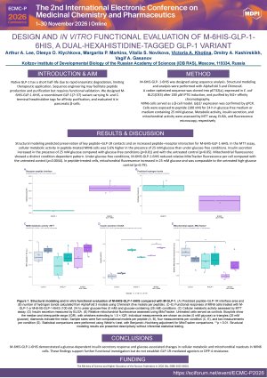
Introduction: Native GLP-1 has a short half-life due to rapid enzymatic degradation, limiting therapeutic application. Sequence engineering may facilitate peptide production and purification but requires functional validation. We designed M-6HIS-GLP-1-6HIS, a recombinant GLP-1(7–37) variant carrying N- and C-terminal hexahistidine tags for affinity purification, and evaluated it in pancreatic β-cells.
Methods: M-6HIS-GLP- 1-6HIS was designed using sequence analysis. Structural modeling and analysis were performed with AlphaFold 3 and ChimeraX. A codon-optimized sequence was cloned into pET32(a), expressed in E. coli BL21(DE3) after 200 µM IPTG induction, and purified by Ni2+-affinity chromatography. MIN6 cells served as a β-cell model. Glp1r expression was confirmed by qPCR. Cells were exposed to peptide (100 nM) for 24 h in glucose-free medium or medium containing 25 mM glucose. Metabolic activity, insulin secretion, and mitochondrial activity were assessed by MTT assay, ELISA, and fluorescence microscopy, respectively.
Results: Structural modeling predicted preservation of key peptide–GLP-1R contacts and an increased peptide–receptor interaction for M-6HIS-GLP-1-6HIS. In the MTT assay, cellular metabolic activity in peptide-treated MIN6 cells was 5.6% higher in the presence of 25 mM glucose than under glucose-free conditions. Insulin secretion increased in the presence of 25 mM glucose compared with glucose-free conditions (p<0.01) and with the untreated control (p<0.05). Mitochondrial fluorescence showed a distinct condition-dependent pattern. Under glucose-free conditions, M-6HIS-GLP-1-6HIS reduced relative MitoTracker fluorescence per cell compared with the untreated control (p=0.0044). In peptide-treated cells, mitochondrial fluorescence increased in 25 mM glucose and was comparable to the untreated high-glucose control (p=0.79).
Conclusions: M-6HIS-GLP-1-6HIS demonstrated a glucose-dependent insulin secretory response and glucose-associated changes in cellular metabolic and mitochondrial readouts in MIN6 cells. These findings support further functional investigation but do not establish GLP-1R-mediated agonism or DPP-4 resistance.
Keywords
GLP-1
recombinant peptide
peptide engineering
MIN6 cells
insulin secretion
hexahistidine tag
Poster
3.pdf
Effects of Morus alba L. and Morus nigra L. Extracts on Memory Performance in a Scopolamine-induced Zebrafish Model
Antidiabetic properties of methanol extract of Dacryodes klaineana leaf in alloxan-induced diabetic rats