Event submissions
Published
This submission belongs to the session S4. Behavioral Ecology of Aquatic Animals: Implications for Habitat Conservation and Ecosystem Services of the event The 1st International Online Conference on Fishes
Published date
03 Nov, 2026
Academic Editor
 Maria Angeles Esteban
Citation
Luisa Araujo Alves Silva, Fabio Aremil Costa dos Santos, Alessandro Loureiro Paschoalini, André de Sena Souza, Wagner Junio de Freitas Martins, Rafael José Furtado de Souza, Gisele Cristina Favero, Nilo Brazzoli, Ronald Kennedy Luz, Gill histopathological responses of Amphilophus citrinellus larvae subjected to different salinities during larviculture, in Proceedings of The 1st International Online Conference on Fishes, 5 November–6 November 2026, MDPI: Basel, Switzerland
Share
Email
Facebook
Twitter
LinkedIn
Gill histopathological responses of Amphilophus citrinellus larvae subjected to different salinities during larviculture

Luisa Araujo Alves Silva 1

Fabio Aremil Costa dos Santos 1
Alessandro Loureiro Paschoalini 2
André de Sena Souza 1
Wagner Junio de Freitas Martins 1
Rafael José Furtado de Souza 1

Gisele Cristina Favero 1
Nilo Brazzoli 2

Ronald Kennedy Luz 1
1. Aquaculture Laboratory, Department of Animal Science, Federal University of Minas Gerais (UFMG), 31270-901 Belo Horizonte, Minas Gerais, Brazil
2. Graduate Program in Biodiversity and Environment, Pontifical Catholic University of Minas Gerais (PUC Minas), Av. Dom José Gaspar, 500, Coração Eucarístico, Building 41, Belo Horizonte, MG, Brazil
Abstract
Introduction:
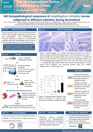
Gills are essential organs in fish, performing functions such as gas exchange, nitrogenous waste excretion, and osmoregulation. Their morphological and functional plasticity in response to environmental variations makes gill histopathology a useful indicator of physiological responses to environmental stress. This study aimed to describe gill histopathological alterations in Amphilophus citrinellus larvae exposed to different salinities during larviculture.
Methods:
Larvae at 5 days of age were reared for 30 days at salinities of 0, 2, 4, 6, 8, and 10 g salt L⁻¹, with four replicates per treatment and 14 larvae per replicate. During the first 15 days, larvae were fed Artemia sp. nauplii and subsequently received a commercial diet containing 450.0 g kg⁻¹ crude protein. At the end of the experiment, gills were collected, histologically processed, and evaluated. Four slides per treatment were selected, with three photomicrographs analyzed per slide. Histopathological alterations were quantified using the histopathological alteration index (HAI), and lesions were classified according to the degree of tissue impairment. Data were analyzed using the Kruskal–Wallis test followed by Dunn's test.
Results:
Larvae maintained at 0 g salt L⁻¹ exhibited normal gill organization, with preserved primary and secondary lamellae and epithelial integrity. At 2 and 4 g salt L⁻¹, edema in the secondary lamellae and mucous cell hyperplasia were observed, indicating an adaptive response. At 6 and 8 g salt L⁻¹, congestion of the central venous sinus was also observed, suggesting vascular impairment. Exposure to 10 g salt L⁻¹ caused structural damage, including disorganization and fusion of secondary lamellae and aneurysm formation.
Conclusion:
Increasing salinity promoted progressive gill histopathological alterations in A. citrinellus, ranging from adaptive responses at low salinities to structural damage under hypersaline conditions, demonstrating the sensitivity of gill tissue to osmotic stress during early development.
Keywords
Amphilophus citrinellus
larviculture
gill histopathology
osmotic stress
salinity tolerance
ornamental aquaculture.
Poster
Poster IOCF_LuisaAASilva.pdf
Abundance, Distribution and Diversity of Macrobenthos in Chittagong Coast, Bangladesh
Fish Diversity and Community Composition of the Xijiang River (Wuzhou Section) in Guangxi, China