Event submissions
Published

This submission belongs to the session 03. Biochemical sensors and assays of the event The 7th International Multidisciplinary Conference on Optofluidics 2017
Published date
21 Jul, 2017
Citation
Li Zhang, Well-ordered plasmonic nanostructure for biosensing, in Proceedings of The 7th International Multidisciplinary Conference on Optofluidics 2017, Singapore, 25 July–28 July 2017, MDPI: Basel, Switzerland, doi: 10.3390/optofluidics2017-04214
Share
Email
Facebook
Twitter
LinkedIn
Well-ordered plasmonic nanostructure for biosensing
'%3e%3cpath%20d='M12.6647%2010.9104C12.4176%2010.325%2012.0589%209.7933%2011.6088%209.34485C11.16%208.89511%2010.6283%208.53653%2010.0432%208.28891C10.038%208.28629%2010.0327%208.28498%2010.0275%208.28236C10.8437%207.69282%2011.3743%206.73252%2011.3743%205.64907C11.3743%203.85423%209.92005%202.40002%208.12522%202.40002C6.33038%202.40002%204.87618%203.85423%204.87618%205.64907C4.87618%206.73252%205.40677%207.69282%206.22296%208.28367C6.21772%208.28629%206.21248%208.2876%206.20724%208.29022C5.62031%208.53783%205.09365%208.89287%204.64167%209.34616C4.19192%209.79496%203.83334%2010.3266%203.58573%2010.9117C3.34247%2011.4846%203.21128%2012.0987%203.19925%2012.721C3.1989%2012.735%203.20135%2012.7489%203.20646%2012.7619C3.21157%2012.7749%203.21924%2012.7868%203.22901%2012.7968C3.23877%2012.8068%203.25045%2012.8148%203.26334%2012.8202C3.27623%2012.8256%203.29007%2012.8284%203.30406%2012.8284H4.09012C4.14776%2012.8284%204.19362%2012.7825%204.19493%2012.7262C4.22113%2011.7148%204.62726%2010.7676%205.34519%2010.0497C6.08802%209.30686%207.07452%208.89811%208.12522%208.89811C9.17592%208.89811%2010.1624%209.30686%2010.9052%2010.0497C11.6232%2010.7676%2012.0293%2011.7148%2012.0555%2012.7262C12.0568%2012.7839%2012.1027%2012.8284%2012.1603%2012.8284H12.9464C12.9604%2012.8284%2012.9742%2012.8256%2012.9871%2012.8202C13%2012.8148%2013.0117%2012.8068%2013.0214%2012.7968C13.0312%2012.7868%2013.0389%2012.7749%2013.044%2012.7619C13.0491%2012.7489%2013.0515%2012.735%2013.0512%2012.721C13.0381%2012.0947%2012.9084%2011.4856%2012.6647%2010.9104ZM8.12522%207.90243C7.52388%207.90243%206.95792%207.66793%206.53214%207.24215C6.10636%206.81636%205.87185%206.2504%205.87185%205.64907C5.87185%205.04773%206.10636%204.48177%206.53214%204.05599C6.95792%203.63021%207.52388%203.3957%208.12522%203.3957C8.72655%203.3957%209.29252%203.63021%209.7183%204.05599C10.1441%204.48177%2010.3786%205.04773%2010.3786%205.64907C10.3786%206.2504%2010.1441%206.81636%209.7183%207.24215C9.29252%207.66793%208.72655%207.90243%208.12522%207.90243Z'%20fill='%235D1EE1'/%3e%3c/g%3e%3c/svg%3e)
Li Zhang 1
1. School of Engineering, Sun Yat-Sen University, China
Abstract
An LSPR biosensor holds attractive advantages of low-cost, capacity of high integration, requirement of simple optical configuration, surpassing commercial mechanical, or propagating surface plasmon resonance (PSPR) biosensors for construction of miniaturized biomedical devices. Well-designed metal nanostructures with “hot-spots” (the region with intense electromagnetic field, mostly located at sharp nanotips, nanoedges, or nanoslits), possessing superior bulk refractometric and molecular detection sensitivity, are ideal candidates for LSPR biosensors. Therefore, sharp tips in nanostructures play an essential role in improving the performance of LSPR sensor.
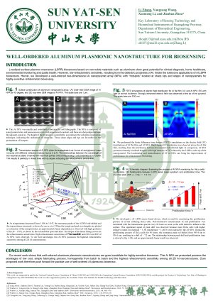
Here, we present an overview of our recent work on using a well-ordered plasmonic nanopyramid array with sharp tips for highly-sensitive refractometric and surface enhanced Raman biosensing. Firstly, we introduce a well-ordered Al nanopyramid array (NPA), fabricated by a facile method of elastic soft lithography and subsequent metal deposition. This Al NPA with sharp tips possess high refractometric sensitivity of ~ 820.4 nm/RIU, which even exceeds that of metal nanobranches. Secondly, we characterize the strongly enhanced local electric field at the tip of nanopyramid in comparison to Al flat film (FF), by performing finite difference time domain (FDTD) simulations. Lastly, we apply this plasmonic nanostructure to real-time monitoring the proliferation of Hella cells. The peak positions of the measured transmission spectra is found linear to the culturing time. After 96 hours, the maximal peak shift is ~ 7.2 nm.
Keywords
Localized surface plasmon resonance biosensor
hot-spot
refractometric biosensing
plasmonic
Poster
POSTER_OPTOFLUIDICS2017_LI ZHANG.pdf
QUANTITATIVE PATHOGEN DETECTION BASED ON DIGITAL LOOP-MEDIATED ISOTHERMAL AMPLIFICATION USING CROSS-INTERFACE EMULSIFICATION TECHNOLOGY
A portable image-based cytometer for rapid malaria detection and quantification