Event submissions
EventsThe 1st International Electronic Conference on Cancers: Exploiting Cancer Vulnerability by Targeting the DNA Damage Response
Published

This submission belongs to the session 8. Poster of the event The 1st International Electronic Conference on Cancers: Exploiting Cancer Vulnerability by Targeting the DNA Damage Response
Published date
29 Jan, 2021
Citation
Hannah Louise Smith, Asima Mukhopadhyay, Yvette Drew, Elaine Willmore, Nicola Curtin, Differences in durability of PARP inhibition by PARP inhibitors in ovarian cancer cells, in Proceedings of The 1st International Electronic Conference on Cancers: Exploiting Cancer Vulnerability by Targeting the DNA Damage Response, 1 February–14 February 2021, MDPI: Basel, Switzerland, doi: 10.3390/IECC2021-09194
Share
Email
Facebook
Twitter
LinkedIn
Differences in durability of PARP inhibition by PARP inhibitors in ovarian cancer cells
'%3e%3cpath%20d='M12.6647%2010.9104C12.4176%2010.325%2012.0589%209.7933%2011.6088%209.34485C11.16%208.89511%2010.6283%208.53653%2010.0432%208.28891C10.038%208.28629%2010.0327%208.28498%2010.0275%208.28236C10.8437%207.69282%2011.3743%206.73252%2011.3743%205.64907C11.3743%203.85423%209.92005%202.40002%208.12522%202.40002C6.33038%202.40002%204.87618%203.85423%204.87618%205.64907C4.87618%206.73252%205.40677%207.69282%206.22296%208.28367C6.21772%208.28629%206.21248%208.2876%206.20724%208.29022C5.62031%208.53783%205.09365%208.89287%204.64167%209.34616C4.19192%209.79496%203.83334%2010.3266%203.58573%2010.9117C3.34247%2011.4846%203.21128%2012.0987%203.19925%2012.721C3.1989%2012.735%203.20135%2012.7489%203.20646%2012.7619C3.21157%2012.7749%203.21924%2012.7868%203.22901%2012.7968C3.23877%2012.8068%203.25045%2012.8148%203.26334%2012.8202C3.27623%2012.8256%203.29007%2012.8284%203.30406%2012.8284H4.09012C4.14776%2012.8284%204.19362%2012.7825%204.19493%2012.7262C4.22113%2011.7148%204.62726%2010.7676%205.34519%2010.0497C6.08802%209.30686%207.07452%208.89811%208.12522%208.89811C9.17592%208.89811%2010.1624%209.30686%2010.9052%2010.0497C11.6232%2010.7676%2012.0293%2011.7148%2012.0555%2012.7262C12.0568%2012.7839%2012.1027%2012.8284%2012.1603%2012.8284H12.9464C12.9604%2012.8284%2012.9742%2012.8256%2012.9871%2012.8202C13%2012.8148%2013.0117%2012.8068%2013.0214%2012.7968C13.0312%2012.7868%2013.0389%2012.7749%2013.044%2012.7619C13.0491%2012.7489%2013.0515%2012.735%2013.0512%2012.721C13.0381%2012.0947%2012.9084%2011.4856%2012.6647%2010.9104ZM8.12522%207.90243C7.52388%207.90243%206.95792%207.66793%206.53214%207.24215C6.10636%206.81636%205.87185%206.2504%205.87185%205.64907C5.87185%205.04773%206.10636%204.48177%206.53214%204.05599C6.95792%203.63021%207.52388%203.3957%208.12522%203.3957C8.72655%203.3957%209.29252%203.63021%209.7183%204.05599C10.1441%204.48177%2010.3786%205.04773%2010.3786%205.64907C10.3786%206.2504%2010.1441%206.81636%209.7183%207.24215C9.29252%207.66793%208.72655%207.90243%208.12522%207.90243Z'%20fill='%235D1EE1'/%3e%3c/g%3e%3c/svg%3e)
Hannah Louise Smith 1
Asima Mukhopadhyay 1,2
Yvette Drew 1,3
Elaine Willmore 1
Nicola Curtin 1
1. Newcastle University, Newcastle upon Tyne, Tyne and Wear, NE1 7RU, United Kingdom.
2. Northern Gynecological Oncology Centre, Queen Elizabeth Hospital, Sherriff Hill, Gateshead NE9 6SX, UK
3. Northern Centre for Cancer Care, The Newcastle upon Tyne Hospitals NHS Foundation Trust, Freeman Hospital, Newcastle upon Tyne NE7 7DN, United Kingdom
Abstract
Background:
PARP inhibitors (PARPi) exploit defects in homologous recombination repair (HRR) to selectively kill tumour cells. Continuous, PARP inhibition is required for cytotoxicity. PARPis rucaparib, olaparib and niraparib have been approved for use in ovarian cancer on continuous schedules. Previous studies demonstrate prolonged PARP inhibition by rucaparb1.
Aim:
To determine if persistent PARP inhibition is a class effect.
Methods:
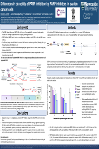
IGROV-1 (human ovarian cancer) cells were treated with 1µM of rucaparib, olaparib, niraparib, talazoparib or pamiparib for 1h before drug was washed off and replaced with fresh media for 0, 1, 24, 48 or 72h prior to harvesting. Cellular PARP activity was measured using a GCLP-validated assay2 in comparison with untreated controls and where 1µM inhibitor was added to the reaction.
Results:
Rucaparib, olaparib, niraparib, talazoparib and pamiparib each inhibited PARP activity in permeabilised cells >99% when 1µM was present during the reaction. After 2 h in drug-free medium rucaparib-induced PARP inhibition was maintained at 92.3 ± 4.3% but was much less with talazoparib (58.6 ±5.0%), pamiparib (56.0 ± 4.5%) olaparib (48.3 ± 19.8%) and niraparib (37.3 ± 11.6%). PARP inhibition in rucaparib-treated cells was maintained for 72h in drug-free medium (77.7 ± 12.3%). This sustained PARP inhibition was not observed with the other PARPis. PARP inhibition was only 12.3 ± 5.2% and 12.5 ± 4.9% 72h after talazoparib and pamiparib, respectively, and undetectable with olaparib and niraparib.
Conclusion:
Rucaparib is unique in its ability to cause persistent PARP inhibition and it is not a class effect. These data have clinical implications for the different uses of PARPi, as a single agent use to exploit HRR defects versus chemo- and radiosensitisation.
1 Murray, J.; et al. BJC, 110, 1977-1984 (2014)
2 Plummer ER, et al Clin Cancer Res. 11 3402-3409 (2005)
Keywords
PARP-1
ovarian cancer
Manuscript
iecc2021 poster final .pdf
Poster
IECC poster edited 03.02.21 PDF.pdf
The roles of cell cycle and BRCA1 in the DNA damage response
Identification of genetic determinants of DNA mismatch repair loss that predict response to immune checkpoint blockade