Event submissions
EventsMicromachines 2021 — 1st International Conference on Micromachines and Applications (ICMA2021)
Published

This submission belongs to the session S3. Micromachines for bio-medical applications of the event Micromachines 2021 — 1st International Conference on Micromachines and Applications (ICMA2021)
Published date
16 Apr, 2021
Citation
Inês Maia Gonçalves, Miguel Madureira, Inês Miranda, Helmut Schütte, Ana Moita, Graça Minas, Stefan Gassmann, Rui Lima, Separation microfluidic device fabricated by micromilling techniques, in Proceedings of Micromachines 2021 — 1st International Conference on Micromachines and Applications (ICMA2021), 15 April–30 April 2021, MDPI: Basel, Switzerland, doi: 10.3390/Micromachines2021-09599
Share
Email
Facebook
Twitter
LinkedIn
Separation microfluidic device fabricated by micromilling techniques
'%3e%3cpath%20d='M12.6647%2010.9104C12.4176%2010.325%2012.0589%209.7933%2011.6088%209.34485C11.16%208.89511%2010.6283%208.53653%2010.0432%208.28891C10.038%208.28629%2010.0327%208.28498%2010.0275%208.28236C10.8437%207.69282%2011.3743%206.73252%2011.3743%205.64907C11.3743%203.85423%209.92005%202.40002%208.12522%202.40002C6.33038%202.40002%204.87618%203.85423%204.87618%205.64907C4.87618%206.73252%205.40677%207.69282%206.22296%208.28367C6.21772%208.28629%206.21248%208.2876%206.20724%208.29022C5.62031%208.53783%205.09365%208.89287%204.64167%209.34616C4.19192%209.79496%203.83334%2010.3266%203.58573%2010.9117C3.34247%2011.4846%203.21128%2012.0987%203.19925%2012.721C3.1989%2012.735%203.20135%2012.7489%203.20646%2012.7619C3.21157%2012.7749%203.21924%2012.7868%203.22901%2012.7968C3.23877%2012.8068%203.25045%2012.8148%203.26334%2012.8202C3.27623%2012.8256%203.29007%2012.8284%203.30406%2012.8284H4.09012C4.14776%2012.8284%204.19362%2012.7825%204.19493%2012.7262C4.22113%2011.7148%204.62726%2010.7676%205.34519%2010.0497C6.08802%209.30686%207.07452%208.89811%208.12522%208.89811C9.17592%208.89811%2010.1624%209.30686%2010.9052%2010.0497C11.6232%2010.7676%2012.0293%2011.7148%2012.0555%2012.7262C12.0568%2012.7839%2012.1027%2012.8284%2012.1603%2012.8284H12.9464C12.9604%2012.8284%2012.9742%2012.8256%2012.9871%2012.8202C13%2012.8148%2013.0117%2012.8068%2013.0214%2012.7968C13.0312%2012.7868%2013.0389%2012.7749%2013.044%2012.7619C13.0491%2012.7489%2013.0515%2012.735%2013.0512%2012.721C13.0381%2012.0947%2012.9084%2011.4856%2012.6647%2010.9104ZM8.12522%207.90243C7.52388%207.90243%206.95792%207.66793%206.53214%207.24215C6.10636%206.81636%205.87185%206.2504%205.87185%205.64907C5.87185%205.04773%206.10636%204.48177%206.53214%204.05599C6.95792%203.63021%207.52388%203.3957%208.12522%203.3957C8.72655%203.3957%209.29252%203.63021%209.7183%204.05599C10.1441%204.48177%2010.3786%205.04773%2010.3786%205.64907C10.3786%206.2504%2010.1441%206.81636%209.7183%207.24215C9.29252%207.66793%208.72655%207.90243%208.12522%207.90243Z'%20fill='%235D1EE1'/%3e%3c/g%3e%3c/svg%3e)
Inês Maia Gonçalves 1,2
Miguel Madureira 3
Inês Miranda 4
Helmut Schütte 3

Ana Moita 2,5

Graça Minas 4

Stefan Gassmann 3

Rui Lima 1,6
1. METRICS, University of Minho, Guimarães, Portugal
2. Instituto Superior Técnico, Universidade de Lisboa, Lisboa, Portugal
3. Jade University of Applied Science, Wilhelmshaven, Germany
4. Center for MicroElectromechanical Systems (CMEMS-UMinho), University of Minho, Guimarães, Portugal
5. CINAMIL, Military Academy Research Center, Portuguese Military Academy, Lisbon, Portugal
6. CEFT, Faculty of Engineering of the University of Porto, Porto, Portugal
Abstract
The diagnostic of several diseases can be performed by analyzing the blood plasma of the patient. Despite the extensive research work, there is still the need to improve the current low-cost fabrication techniques and devices for the separation of the plasma from the blood cells. Microfluidic biomedical devices have a great potential for that process. Hence, a microfluidic device made by micromilling and sealed with the oxygen plasma technique was tested by means of two different blood analogue fluids. The device has four microchannels having similar geometries but with different channel depths. A high-speed video microscopy system was used for the visualization and acquisition of the flow of the analogue fluids throughout the microchannels of the device. Then, the separation of particles and plasma was evaluated using the software ImageJ by measuring and comparing the grey values at the entrance and at the exit of the channel. The device has shown a significant reduction of the amount of cells between the entrance and the exit of the microchannels. No major influences were found from the depth of the channels and size of the particles on the separation process. However, it was found that flow rate has affected the separation results where the best results were obtained for a flow rate of 100 μL/min. Although these results are promising, further analysis and optimizations of the microfluidic devices will be conducted in future works, as well as, the comparison between devices sealed using different methods, such as, the solvent bonding technique.
Keywords
Micromilling
microfluidic devices
particle separation
blood analogues
image analysis
Poster
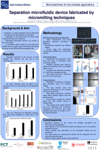
ICMA2021_Separation_Poster.pdf
Stable Porous Silicon Membranes for Fast Bacterial Detection
Rapid lipid content screening in Neochloris Oleoabundans by carbon-based dielectrophoresis