Event submissions
Published
This submission belongs to the session S1. Dental Biomaterials of the event The 2nd International Online Conference on Functional Biomaterials
Published date
03 Jul, 2026
Academic Editor
'%3e%3cpath%20d='M12.6657%2010.9104C12.4185%2010.325%2012.0599%209.79327%2011.6097%209.34482C11.1609%208.89508%2010.6293%208.5365%2010.0442%208.28888C10.0389%208.28626%2010.0337%208.28495%2010.0285%208.28233C10.8446%207.69279%2011.3752%206.73249%2011.3752%205.64904C11.3752%203.8542%209.92103%202.39999%208.1262%202.39999C6.33136%202.39999%204.87715%203.8542%204.87715%205.64904C4.87715%206.73249%205.40774%207.69279%206.22393%208.28364C6.21869%208.28626%206.21345%208.28757%206.20821%208.29019C5.62129%208.5378%205.09463%208.89284%204.64265%209.34613C4.1929%209.79493%203.83432%2010.3266%203.58671%2010.9117C3.34345%2011.4845%203.21226%2012.0987%203.20023%2012.7209C3.19988%2012.7349%203.20233%2012.7488%203.20744%2012.7619C3.21255%2012.7749%203.22022%2012.7867%203.22998%2012.7968C3.23975%2012.8068%203.25142%2012.8147%203.26431%2012.8202C3.2772%2012.8256%203.29105%2012.8284%203.30504%2012.8284H4.09109C4.14874%2012.8284%204.19459%2012.7825%204.1959%2012.7262C4.2221%2011.7148%204.62823%2010.7676%205.34617%2010.0497C6.08899%209.30683%207.0755%208.89808%208.1262%208.89808C9.17689%208.89808%2010.1634%209.30683%2010.9062%2010.0497C11.6242%2010.7676%2012.0303%2011.7148%2012.0565%2012.7262C12.0578%2012.7838%2012.1037%2012.8284%2012.1613%2012.8284H12.9474C12.9613%2012.8284%2012.9752%2012.8256%2012.9881%2012.8202C13.001%2012.8147%2013.0126%2012.8068%2013.0224%2012.7968C13.0322%2012.7867%2013.0398%2012.7749%2013.0449%2012.7619C13.0501%2012.7488%2013.0525%2012.7349%2013.0522%2012.7209C13.0391%2012.0947%2012.9094%2011.4855%2012.6657%2010.9104ZM8.1262%207.9024C7.52486%207.9024%206.9589%207.6679%206.53312%207.24211C6.10733%206.81633%205.87283%206.25037%205.87283%205.64904C5.87283%205.0477%206.10733%204.48174%206.53312%204.05596C6.9589%203.63018%207.52486%203.39567%208.1262%203.39567C8.72753%203.39567%209.29349%203.63018%209.71927%204.05596C10.1451%204.48174%2010.3796%205.0477%2010.3796%205.64904C10.3796%206.25037%2010.1451%206.81633%209.71927%207.24211C9.29349%207.6679%208.72753%207.9024%208.1262%207.9024Z'%20fill='%235D1EE1'/%3e%3c/g%3e%3c/svg%3e) Gianrico Spagnuolo
Gianrico SpagnuoloCitation
Niki Karipidou, Eleni Manolakaki, Grigoris Petkoglou, Nikolaos Michailidis, Petros T. Koidis, Amalia Aggeli, Apostolos Argyros, Design and Physical Characterization of 3D-Printed Collagen Scaffolds for Regenerative Dentistry, in Proceedings of The 2nd International Online Conference on Functional Biomaterials, 8 July–10 July 2026, MDPI: Basel, Switzerland
Share
Email
Facebook
Twitter
LinkedIn
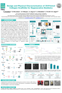
Design and Physical Characterization of 3D-Printed Collagen Scaffolds for Regenerative Dentistry
'%3e%3cpath%20d='M12.6647%2010.9104C12.4176%2010.325%2012.0589%209.7933%2011.6088%209.34485C11.16%208.89511%2010.6283%208.53653%2010.0432%208.28891C10.038%208.28629%2010.0327%208.28498%2010.0275%208.28236C10.8437%207.69282%2011.3743%206.73252%2011.3743%205.64907C11.3743%203.85423%209.92005%202.40002%208.12522%202.40002C6.33038%202.40002%204.87618%203.85423%204.87618%205.64907C4.87618%206.73252%205.40677%207.69282%206.22296%208.28367C6.21772%208.28629%206.21248%208.2876%206.20724%208.29022C5.62031%208.53783%205.09365%208.89287%204.64167%209.34616C4.19192%209.79496%203.83334%2010.3266%203.58573%2010.9117C3.34247%2011.4846%203.21128%2012.0987%203.19925%2012.721C3.1989%2012.735%203.20135%2012.7489%203.20646%2012.7619C3.21157%2012.7749%203.21924%2012.7868%203.22901%2012.7968C3.23877%2012.8068%203.25045%2012.8148%203.26334%2012.8202C3.27623%2012.8256%203.29007%2012.8284%203.30406%2012.8284H4.09012C4.14776%2012.8284%204.19362%2012.7825%204.19493%2012.7262C4.22113%2011.7148%204.62726%2010.7676%205.34519%2010.0497C6.08802%209.30686%207.07452%208.89811%208.12522%208.89811C9.17592%208.89811%2010.1624%209.30686%2010.9052%2010.0497C11.6232%2010.7676%2012.0293%2011.7148%2012.0555%2012.7262C12.0568%2012.7839%2012.1027%2012.8284%2012.1603%2012.8284H12.9464C12.9604%2012.8284%2012.9742%2012.8256%2012.9871%2012.8202C13%2012.8148%2013.0117%2012.8068%2013.0214%2012.7968C13.0312%2012.7868%2013.0389%2012.7749%2013.044%2012.7619C13.0491%2012.7489%2013.0515%2012.735%2013.0512%2012.721C13.0381%2012.0947%2012.9084%2011.4856%2012.6647%2010.9104ZM8.12522%207.90243C7.52388%207.90243%206.95792%207.66793%206.53214%207.24215C6.10636%206.81636%205.87185%206.2504%205.87185%205.64907C5.87185%205.04773%206.10636%204.48177%206.53214%204.05599C6.95792%203.63021%207.52388%203.3957%208.12522%203.3957C8.72655%203.3957%209.29252%203.63021%209.7183%204.05599C10.1441%204.48177%2010.3786%205.04773%2010.3786%205.64907C10.3786%206.2504%2010.1441%206.81636%209.7183%207.24215C9.29252%207.66793%208.72655%207.90243%208.12522%207.90243Z'%20fill='%235D1EE1'/%3e%3c/g%3e%3c/svg%3e)
Niki Karipidou 1,2
Eleni Manolakaki 1
Grigoris Petkoglou 1
Apostolos Argyros 2,3

Nikolaos Michailidis 2,3
Petros T. Koidis 4
Amalia Aggeli 1,2
1. Dpt of Chemical Engineering, Faculty of Engineering, Aristotle University of Thessaloniki, 54124 Thessaloniki, Greece, Greece
2. Centre for Research & Development of Advanced Materials (CERDAM), Center for Interdisciplinary Research and Innovation, Balkan Center, 57001 Thessaloniki, Greece
3. Department of Mechanical Engineering, Faculty of Engineering, Aristotle University of Thessaloniki, 54124 Thessaloniki, Greece, Greece
4. Dpt of Prosthodontics, School of Dentistry, Faculty of Health Sciences, Aristotle University of Thessaloniki, 54124 Thessaloniki, Greece, Greece
Abstract
Tissue engineering in regenerative dentistry integrates biomaterials and biofabrication strategies to develop advanced solutions for oral tissue repair; however, significant material and structural challenges persist due to the heterogeneous nature of oral tissues and the limited regenerative capacity of alveolar bone. Additive manufacturing offers a promising alternative to conventional grafting approaches, which are frequently associated with donor site morbidity, immune reactions, and clinical complications. In this study, collagen-based biomaterial systems were developed and systematically evaluated for extrusion-based three-dimensional (3D) bioprinting of scaffolds intended for alveolar bone regeneration. Four fabrication protocols were comparatively assessed, including a standard and optimized protocol, an externally crosslinked system, and a cost-effective approach employing a conventional 3D printer, enabling evaluation of processing–structure–property relationships. Following extrusion and freeze-drying, scaffold performance was characterized through comprehensive rheological analyses, as well as morphological and thermal evaluation using scanning electron microscopy (SEM), differential scanning calorimetry, and quantitative pore size analysis. Rheological results demonstrated consistent viscoelastic and shear-thinning behavior across all formulations, with well-defined linear viscoelastic regions and stable elastic responses over wide frequency ranges, indicating suitability for extrusion-based processing and post-fabrication integrity. SEM analysis revealed protocol-dependent microarchitectures, ranging from highly interconnected micro- and macro-porous networks favorable for mass transport to denser extracellular matrix-mimicking collagen fiber arrangements governed by a crosslinking strategy. Thermal analysis confirmed amorphous scaffold structures with distinct energy absorption profiles reflecting variations in network stability. While all protocols demonstrated potential for bone regeneration, the optimized standard protocol exhibited comparatively enhanced viscoelastic stability, interconnected porosity, and thermal resistance. Bioprinting results further demonstrate scaffold printability and provide insight into current processing limitations and directions for optimization. These findings highlight the critical role of bioink formulation and scaffold design in achieving mechanically robust, biomimetic structures, and support the continued development of 3D-printed collagen scaffolds as promising candidates for regenerative dentistry applications.
Keywords
collagen scaffolds
regenerative dentistry
3D bioprinting
biomimetic structures
Poster
Karipidou_Niki_IOCFB2026_Poster Presentation_Dental_Biomaterials.pdf
Graphene Oxide-Modified Portland Cement Enhances Bioactivity and Periapical Healing: An In Vitro and In Vivo Study
Microstructurally Refined Ti-6Al-7Nb via High-Pressure Sliding Modulates Osteoblast Adhesion and Proliferation