Event submissions
Published
This submission belongs to the session S2. Bone Biomaterials of the event The 2nd International Online Conference on Functional Biomaterials
Published date
03 Jul, 2026
Academic Editor
'%3e%3cpath%20d='M12.6657%2010.9104C12.4185%2010.325%2012.0599%209.79327%2011.6097%209.34482C11.1609%208.89508%2010.6293%208.5365%2010.0442%208.28888C10.0389%208.28626%2010.0337%208.28495%2010.0285%208.28233C10.8446%207.69279%2011.3752%206.73249%2011.3752%205.64904C11.3752%203.8542%209.92103%202.39999%208.1262%202.39999C6.33136%202.39999%204.87715%203.8542%204.87715%205.64904C4.87715%206.73249%205.40774%207.69279%206.22393%208.28364C6.21869%208.28626%206.21345%208.28757%206.20821%208.29019C5.62129%208.5378%205.09463%208.89284%204.64265%209.34613C4.1929%209.79493%203.83432%2010.3266%203.58671%2010.9117C3.34345%2011.4845%203.21226%2012.0987%203.20023%2012.7209C3.19988%2012.7349%203.20233%2012.7488%203.20744%2012.7619C3.21255%2012.7749%203.22022%2012.7867%203.22998%2012.7968C3.23975%2012.8068%203.25142%2012.8147%203.26431%2012.8202C3.2772%2012.8256%203.29105%2012.8284%203.30504%2012.8284H4.09109C4.14874%2012.8284%204.19459%2012.7825%204.1959%2012.7262C4.2221%2011.7148%204.62823%2010.7676%205.34617%2010.0497C6.08899%209.30683%207.0755%208.89808%208.1262%208.89808C9.17689%208.89808%2010.1634%209.30683%2010.9062%2010.0497C11.6242%2010.7676%2012.0303%2011.7148%2012.0565%2012.7262C12.0578%2012.7838%2012.1037%2012.8284%2012.1613%2012.8284H12.9474C12.9613%2012.8284%2012.9752%2012.8256%2012.9881%2012.8202C13.001%2012.8147%2013.0126%2012.8068%2013.0224%2012.7968C13.0322%2012.7867%2013.0398%2012.7749%2013.0449%2012.7619C13.0501%2012.7488%2013.0525%2012.7349%2013.0522%2012.7209C13.0391%2012.0947%2012.9094%2011.4855%2012.6657%2010.9104ZM8.1262%207.9024C7.52486%207.9024%206.9589%207.6679%206.53312%207.24211C6.10733%206.81633%205.87283%206.25037%205.87283%205.64904C5.87283%205.0477%206.10733%204.48174%206.53312%204.05596C6.9589%203.63018%207.52486%203.39567%208.1262%203.39567C8.72753%203.39567%209.29349%203.63018%209.71927%204.05596C10.1451%204.48174%2010.3796%205.0477%2010.3796%205.64904C10.3796%206.25037%2010.1451%206.81633%209.71927%207.24211C9.29349%207.6679%208.72753%207.9024%208.1262%207.9024Z'%20fill='%235D1EE1'/%3e%3c/g%3e%3c/svg%3e) Elisa Boanini
Elisa BoaniniCitation
CRISTIAN ENRIQUE TORRES-SALCIDO, Aída Gutiérrez-Alejandre, Marco Antonio Álvarez-Pérez, Jesús Ángel Arenas-Alatorre, Vincenzo Guarino, Janeth Serrano-Bello, Development and biological in vitro study of three-dimensional printed poly(lactic acid) scaffolds coated with gelatin-based coaxial nanofibrous coatings for bone repair, in Proceedings of The 2nd International Online Conference on Functional Biomaterials, 8 July–10 July 2026, MDPI: Basel, Switzerland
Share
Email
Facebook
Twitter
LinkedIn
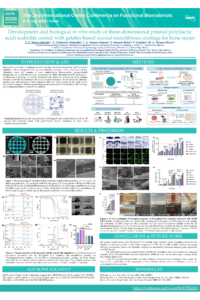
Development and biological in vitro study of three-dimensional printed poly(lactic acid) scaffolds coated with gelatin-based coaxial nanofibrous coatings for bone repair
'%3e%3cpath%20d='M12.6647%2010.9104C12.4176%2010.325%2012.0589%209.7933%2011.6088%209.34485C11.16%208.89511%2010.6283%208.53653%2010.0432%208.28891C10.038%208.28629%2010.0327%208.28498%2010.0275%208.28236C10.8437%207.69282%2011.3743%206.73252%2011.3743%205.64907C11.3743%203.85423%209.92005%202.40002%208.12522%202.40002C6.33038%202.40002%204.87618%203.85423%204.87618%205.64907C4.87618%206.73252%205.40677%207.69282%206.22296%208.28367C6.21772%208.28629%206.21248%208.2876%206.20724%208.29022C5.62031%208.53783%205.09365%208.89287%204.64167%209.34616C4.19192%209.79496%203.83334%2010.3266%203.58573%2010.9117C3.34247%2011.4846%203.21128%2012.0987%203.19925%2012.721C3.1989%2012.735%203.20135%2012.7489%203.20646%2012.7619C3.21157%2012.7749%203.21924%2012.7868%203.22901%2012.7968C3.23877%2012.8068%203.25045%2012.8148%203.26334%2012.8202C3.27623%2012.8256%203.29007%2012.8284%203.30406%2012.8284H4.09012C4.14776%2012.8284%204.19362%2012.7825%204.19493%2012.7262C4.22113%2011.7148%204.62726%2010.7676%205.34519%2010.0497C6.08802%209.30686%207.07452%208.89811%208.12522%208.89811C9.17592%208.89811%2010.1624%209.30686%2010.9052%2010.0497C11.6232%2010.7676%2012.0293%2011.7148%2012.0555%2012.7262C12.0568%2012.7839%2012.1027%2012.8284%2012.1603%2012.8284H12.9464C12.9604%2012.8284%2012.9742%2012.8256%2012.9871%2012.8202C13%2012.8148%2013.0117%2012.8068%2013.0214%2012.7968C13.0312%2012.7868%2013.0389%2012.7749%2013.044%2012.7619C13.0491%2012.7489%2013.0515%2012.735%2013.0512%2012.721C13.0381%2012.0947%2012.9084%2011.4856%2012.6647%2010.9104ZM8.12522%207.90243C7.52388%207.90243%206.95792%207.66793%206.53214%207.24215C6.10636%206.81636%205.87185%206.2504%205.87185%205.64907C5.87185%205.04773%206.10636%204.48177%206.53214%204.05599C6.95792%203.63021%207.52388%203.3957%208.12522%203.3957C8.72655%203.3957%209.29252%203.63021%209.7183%204.05599C10.1441%204.48177%2010.3786%205.04773%2010.3786%205.64907C10.3786%206.2504%2010.1441%206.81636%209.7183%207.24215C9.29252%207.66793%208.72655%207.90243%208.12522%207.90243Z'%20fill='%235D1EE1'/%3e%3c/g%3e%3c/svg%3e)
CRISTIAN ENRIQUE TORRES-SALCIDO 1
Aída Gutiérrez-Alejandre 2
Jesús Ángel Arenas-Alatorre 3
Janeth Serrano-Bello 1

Vincenzo Guarino 4
Marco Antonio Álvarez-Pérez 1
1. Tissue Bioengineering Laboratory, Division of Postgraduate Studies and Research, Faculty of Dentistry, National Autonomous University of Mexico, Ciudad Universitaria, Mexico City 04510, Mexico, Mexico
2. Catalysis Research Unit, Department of Chemical Engineering, Faculty of Chemistry, National Autonomous University of Mexico, Ciudad Universitaria, Mexico City 04510, Mexico, Mexico
3. Laboratory 113 Synthesis of Magnetic Nanomaterials, Department of Condensed Matter Physics, Institute of Physics, National Autonomous University of Mexico, Ciudad Universitaria, Mexico City 04510, Mexico, Mexico
4. Institute of Polymers, Composites and Biomaterials, National Research Council of Italy, Mostra d’Oltremare, Pad.20, V.le J.F. Kennedy 54, 80125 Naples, Italy, Italy
Abstract
Bone grafting has been a challenge in recent decades, primarily because the "gold standard" methods, such as autografts, have low bioavailability and pose a risk to the donor site. Therefore, there still remains a focus ondeveloping biocompatible polymer-based biomaterials as a scaffolds for bone restoration. Three-dimensional (3D) printing is a revolutionary technology for scaffold manufacturing. However, material extrusion printing remains limited by the resolution and smooth surface formation, which affects cell adhesion, proliferation, and subsequent bone restoration. This work focuses on the study of the effect of the biological behavior of gelatin/polyester-based coaxial nanofibrous (CNF) coatings on 3D-printed poly(lactic acid) (PLA) scaffolds. The results of scanning electron microscopy showed a good integration of the 3D-printed scaffolds and the nanofiber coating structures, while transmission electron microscopy revealed the core–shell structure of the electrospun CNF coatings based on PCL/Gt and PLA/Gt. Biological in vitro activity assessment showed that gelatin-coated scaffold-based CNF coatings promoted the cell adhesion and viability of a human fetal osteoblast (hFOB 1.19) cell line at 24 h and 21 days of culture, respectively, compared to the control, uncoated scaffolds, and those coated with pure polymer fibers. The highest alkaline phosphatase activity was observed on day 7, as an early marker for osteogenic differentiation. The formation of calcium phosphate mineral deposits was evident on day 14 for the CNF coatings. In conclusion, these results indicate that modifying the 3D-printed scaffolds with gelatin-based CNF coatings represents a possible application route for 3D-printed biomaterials for the improvement of the biochemical and topographical properties of scaffolds for bone repair. CET-S acknowledges the scholarship support from SECIHTI for his Ph.D. studies (CVU:1009583). The authors are grateful for the financial support given by the DGAPA-UNAM-PAPIIT IN106624, IN218223, and IN202924, and PAIP-FQ 5000-9222 projects.
Keywords
Bone graft
Three-dimensional printing
Electrospinning
Tissue engineering
coatings
nanofibers
Poster
sciforum-175414 - Torres-Salcido CE.pdf
Bisphosphonate Delivery via a pH-Responsive Bone Implant for Accelerated Repair of Osteoporotic Bone Fractures
BMP7-Modified Polycaprolactone Nanofibrous Scaffolds Improve Osteoblast Activity