Event submissions
Published
This submission belongs to the session S3. Antibacterial Biomaterials and Surfaces of the event The 2nd International Online Conference on Functional Biomaterials
Published date
03 Jul, 2026
Academic Editor
'%3e%3cpath%20d='M12.6657%2010.9104C12.4185%2010.325%2012.0599%209.79327%2011.6097%209.34482C11.1609%208.89508%2010.6293%208.5365%2010.0442%208.28888C10.0389%208.28626%2010.0337%208.28495%2010.0285%208.28233C10.8446%207.69279%2011.3752%206.73249%2011.3752%205.64904C11.3752%203.8542%209.92103%202.39999%208.1262%202.39999C6.33136%202.39999%204.87715%203.8542%204.87715%205.64904C4.87715%206.73249%205.40774%207.69279%206.22393%208.28364C6.21869%208.28626%206.21345%208.28757%206.20821%208.29019C5.62129%208.5378%205.09463%208.89284%204.64265%209.34613C4.1929%209.79493%203.83432%2010.3266%203.58671%2010.9117C3.34345%2011.4845%203.21226%2012.0987%203.20023%2012.7209C3.19988%2012.7349%203.20233%2012.7488%203.20744%2012.7619C3.21255%2012.7749%203.22022%2012.7867%203.22998%2012.7968C3.23975%2012.8068%203.25142%2012.8147%203.26431%2012.8202C3.2772%2012.8256%203.29105%2012.8284%203.30504%2012.8284H4.09109C4.14874%2012.8284%204.19459%2012.7825%204.1959%2012.7262C4.2221%2011.7148%204.62823%2010.7676%205.34617%2010.0497C6.08899%209.30683%207.0755%208.89808%208.1262%208.89808C9.17689%208.89808%2010.1634%209.30683%2010.9062%2010.0497C11.6242%2010.7676%2012.0303%2011.7148%2012.0565%2012.7262C12.0578%2012.7838%2012.1037%2012.8284%2012.1613%2012.8284H12.9474C12.9613%2012.8284%2012.9752%2012.8256%2012.9881%2012.8202C13.001%2012.8147%2013.0126%2012.8068%2013.0224%2012.7968C13.0322%2012.7867%2013.0398%2012.7749%2013.0449%2012.7619C13.0501%2012.7488%2013.0525%2012.7349%2013.0522%2012.7209C13.0391%2012.0947%2012.9094%2011.4855%2012.6657%2010.9104ZM8.1262%207.9024C7.52486%207.9024%206.9589%207.6679%206.53312%207.24211C6.10733%206.81633%205.87283%206.25037%205.87283%205.64904C5.87283%205.0477%206.10733%204.48174%206.53312%204.05596C6.9589%203.63018%207.52486%203.39567%208.1262%203.39567C8.72753%203.39567%209.29349%203.63018%209.71927%204.05596C10.1451%204.48174%2010.3796%205.0477%2010.3796%205.64904C10.3796%206.25037%2010.1451%206.81633%209.71927%207.24211C9.29349%207.6679%208.72753%207.9024%208.1262%207.9024Z'%20fill='%235D1EE1'/%3e%3c/g%3e%3c/svg%3e) John Luong
John LuongCitation
Felipe Cordova Lozano, Luis Emilio Toxqui Martínez, Ana Karen Cordova Estrada, Ivanna Gisell Vazquez Casillas, Regina Alexa Gualito Cervantes, Water-Stable Electrospun PVA Nanofibers via Vapor Crosslinking and Controlled In Situ Silver Nanoparticle Incorporation for Antimicrobial Applications, in Proceedings of The 2nd International Online Conference on Functional Biomaterials, 8 July–10 July 2026, MDPI: Basel, Switzerland
Share
Email
Facebook
Twitter
LinkedIn
Water-Stable Electrospun PVA Nanofibers via Vapor Crosslinking and Controlled In Situ Silver Nanoparticle Incorporation for Antimicrobial Applications
'%3e%3cpath%20d='M12.6647%2010.9104C12.4176%2010.325%2012.0589%209.7933%2011.6088%209.34485C11.16%208.89511%2010.6283%208.53653%2010.0432%208.28891C10.038%208.28629%2010.0327%208.28498%2010.0275%208.28236C10.8437%207.69282%2011.3743%206.73252%2011.3743%205.64907C11.3743%203.85423%209.92005%202.40002%208.12522%202.40002C6.33038%202.40002%204.87618%203.85423%204.87618%205.64907C4.87618%206.73252%205.40677%207.69282%206.22296%208.28367C6.21772%208.28629%206.21248%208.2876%206.20724%208.29022C5.62031%208.53783%205.09365%208.89287%204.64167%209.34616C4.19192%209.79496%203.83334%2010.3266%203.58573%2010.9117C3.34247%2011.4846%203.21128%2012.0987%203.19925%2012.721C3.1989%2012.735%203.20135%2012.7489%203.20646%2012.7619C3.21157%2012.7749%203.21924%2012.7868%203.22901%2012.7968C3.23877%2012.8068%203.25045%2012.8148%203.26334%2012.8202C3.27623%2012.8256%203.29007%2012.8284%203.30406%2012.8284H4.09012C4.14776%2012.8284%204.19362%2012.7825%204.19493%2012.7262C4.22113%2011.7148%204.62726%2010.7676%205.34519%2010.0497C6.08802%209.30686%207.07452%208.89811%208.12522%208.89811C9.17592%208.89811%2010.1624%209.30686%2010.9052%2010.0497C11.6232%2010.7676%2012.0293%2011.7148%2012.0555%2012.7262C12.0568%2012.7839%2012.1027%2012.8284%2012.1603%2012.8284H12.9464C12.9604%2012.8284%2012.9742%2012.8256%2012.9871%2012.8202C13%2012.8148%2013.0117%2012.8068%2013.0214%2012.7968C13.0312%2012.7868%2013.0389%2012.7749%2013.044%2012.7619C13.0491%2012.7489%2013.0515%2012.735%2013.0512%2012.721C13.0381%2012.0947%2012.9084%2011.4856%2012.6647%2010.9104ZM8.12522%207.90243C7.52388%207.90243%206.95792%207.66793%206.53214%207.24215C6.10636%206.81636%205.87185%206.2504%205.87185%205.64907C5.87185%205.04773%206.10636%204.48177%206.53214%204.05599C6.95792%203.63021%207.52388%203.3957%208.12522%203.3957C8.72655%203.3957%209.29252%203.63021%209.7183%204.05599C10.1441%204.48177%2010.3786%205.04773%2010.3786%205.64907C10.3786%206.2504%2010.1441%206.81636%209.7183%207.24215C9.29252%207.66793%208.72655%207.90243%208.12522%207.90243Z'%20fill='%235D1EE1'/%3e%3c/g%3e%3c/svg%3e)
Felipe Cordova Lozano 1
Luis Emilio Toxqui Martínez 2
Ana Karen Cordova Estrada 3
Ivanna Gisell Vazquez Casillas 4
Regina Alexa Gualito Cervantes 4
1. Chemical and Biological Department, Science School, Universidad de las Américas Puebla. Ex hacienda Sta. Catarina Mártir S/N. San Andrés Cholula, Puebla. ZIP 72810. Mexico, Mexico
2. Centro de Investigación y Desarrollo Tecnológico en Electroquímica, Parque Tecnológico Querétaro, s/n, San Fandila, ZIP 76703, Pedro Escobedo, Queretaro, Mexico, Mexico
3. Departament of Civil and Engineering, Universidad de las Américas Puebla UDLAP, San Andrés Cholula, Puebla, México, ZIP 72810, Mexico
4. Department of Chemical Engineering, Universidad de las Américas Puebla UDLAP, San Andrés Cholula, Puebla, México, ZIP 72810, Mexico
Abstract
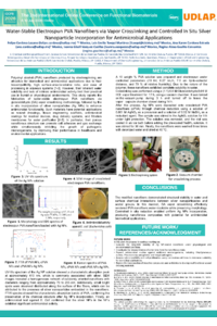
Polyvinyl alcohol (PVA) nanofibers produced by electrospinning are attractive for biomedical and antimicrobial applications due to their biocompatibility and ease of processing in aqueous systems. However, their inherent water solubility and lack of intrinsic antimicrobial activity limit practical use. This study reports the fabrication of water-stable electrospun PVA nanofibers via glutaraldehyde (GA) vapor crosslinking, followed by in situ incorporation of silver nanoparticles (Ag NPs) to enhance antimicrobial functionality. In this study a 10 wt % PVA solution was electrospun under controlled parameters (10 kV, 0.27 mL/h, 7 cm tip-to-collector distance, and 70 % relative humidity). Crosslinking was performed using GA/HCl vapor treatment for 24 h. Ag NPs were deposited onto crosslinked PVA nanofibers (cPVA) through chemical reduction using AgNO₃ as a precursor and NaBH₄ as a reductant agent. Morphological, structural, and chemical characterization was carried out using SEM, EDX, FTIR, and Raman spectroscopy. Nanoparticle size distribution was analyzed with ImageJ software. Electrospun PVA nanofibers exhibited an average diameter of approximately 140 nm, which increased to ~170 nm after crosslinking, confirming successful structural modification. FTIR and Raman spectra verified acetal bridge formation between PVA and GA. Ag NPs with size distributions between 12 and 20 nm were homogeneously deposited using the chemical reduction method, showing reduced agglomeration compared to Tollens-based synthesis. The modified nanofibers demonstrated structural stability in water and surface chemical interactions between silver and acetal groups. In this manner, GA vapor crosslinking effectively rendered PVA nanofibers water-insoluble while preserving morphology. Controlled in situ reduction enabled uniform Ag NP incorporation, producing nanofibrous composites with potential for antimicrobial biomedical applications.
Keywords
PVA nanofibers
Ag nanoparticles
antimicrobial nanomaterials,
Poster
Poster presentations Antimicrobial Properties UDLAP.pdf
Multifunctional Rhenium and Iridium Flavonoid Complexes for Antibacterial Biomaterials and Green Therapeutic Platforms
Balancing Cytocompatibility and Bacterial Resistance: Insights from Silver-Modified Ti6Al4V Co-Culture Studies