Event submissions
Published
This submission belongs to the session S4. Biomaterials for Tissue Engineering and Regenerative Medicine of the event The 2nd International Online Conference on Functional Biomaterials
Published date
03 Jul, 2026
Academic Editor
'%3e%3cpath%20d='M12.6657%2010.9104C12.4185%2010.325%2012.0599%209.79327%2011.6097%209.34482C11.1609%208.89508%2010.6293%208.5365%2010.0442%208.28888C10.0389%208.28626%2010.0337%208.28495%2010.0285%208.28233C10.8446%207.69279%2011.3752%206.73249%2011.3752%205.64904C11.3752%203.8542%209.92103%202.39999%208.1262%202.39999C6.33136%202.39999%204.87715%203.8542%204.87715%205.64904C4.87715%206.73249%205.40774%207.69279%206.22393%208.28364C6.21869%208.28626%206.21345%208.28757%206.20821%208.29019C5.62129%208.5378%205.09463%208.89284%204.64265%209.34613C4.1929%209.79493%203.83432%2010.3266%203.58671%2010.9117C3.34345%2011.4845%203.21226%2012.0987%203.20023%2012.7209C3.19988%2012.7349%203.20233%2012.7488%203.20744%2012.7619C3.21255%2012.7749%203.22022%2012.7867%203.22998%2012.7968C3.23975%2012.8068%203.25142%2012.8147%203.26431%2012.8202C3.2772%2012.8256%203.29105%2012.8284%203.30504%2012.8284H4.09109C4.14874%2012.8284%204.19459%2012.7825%204.1959%2012.7262C4.2221%2011.7148%204.62823%2010.7676%205.34617%2010.0497C6.08899%209.30683%207.0755%208.89808%208.1262%208.89808C9.17689%208.89808%2010.1634%209.30683%2010.9062%2010.0497C11.6242%2010.7676%2012.0303%2011.7148%2012.0565%2012.7262C12.0578%2012.7838%2012.1037%2012.8284%2012.1613%2012.8284H12.9474C12.9613%2012.8284%2012.9752%2012.8256%2012.9881%2012.8202C13.001%2012.8147%2013.0126%2012.8068%2013.0224%2012.7968C13.0322%2012.7867%2013.0398%2012.7749%2013.0449%2012.7619C13.0501%2012.7488%2013.0525%2012.7349%2013.0522%2012.7209C13.0391%2012.0947%2012.9094%2011.4855%2012.6657%2010.9104ZM8.1262%207.9024C7.52486%207.9024%206.9589%207.6679%206.53312%207.24211C6.10733%206.81633%205.87283%206.25037%205.87283%205.64904C5.87283%205.0477%206.10733%204.48174%206.53312%204.05596C6.9589%203.63018%207.52486%203.39567%208.1262%203.39567C8.72753%203.39567%209.29349%203.63018%209.71927%204.05596C10.1451%204.48174%2010.3796%205.0477%2010.3796%205.64904C10.3796%206.25037%2010.1451%206.81633%209.71927%207.24211C9.29349%207.6679%208.72753%207.9024%208.1262%207.9024Z'%20fill='%235D1EE1'/%3e%3c/g%3e%3c/svg%3e) Piergiorgio Gentile
Piergiorgio GentileCitation
Josué Urquiaga Zavaleta, Jeniffer Riva Bocanegra, Raul Siche, Iana Soares Pessoa, Marcio Fronza, Antonio Domingos de Souza Júnior, Gelatin–Chitosan Scaffolds from Mugil cephalus Skin: Enhanced Fibroblast Proliferation, Migration, and Anti-Inflammatory Activity for Wound Healing, in Proceedings of The 2nd International Online Conference on Functional Biomaterials, 8 July–10 July 2026, MDPI: Basel, Switzerland
Share
Email
Facebook
Twitter
LinkedIn
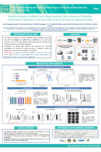
Gelatin–Chitosan Scaffolds from Mugil cephalus Skin: Enhanced Fibroblast Proliferation, Migration, and Anti-Inflammatory Activity for Wound Healing
'%3e%3cpath%20d='M12.6647%2010.9104C12.4176%2010.325%2012.0589%209.7933%2011.6088%209.34485C11.16%208.89511%2010.6283%208.53653%2010.0432%208.28891C10.038%208.28629%2010.0327%208.28498%2010.0275%208.28236C10.8437%207.69282%2011.3743%206.73252%2011.3743%205.64907C11.3743%203.85423%209.92005%202.40002%208.12522%202.40002C6.33038%202.40002%204.87618%203.85423%204.87618%205.64907C4.87618%206.73252%205.40677%207.69282%206.22296%208.28367C6.21772%208.28629%206.21248%208.2876%206.20724%208.29022C5.62031%208.53783%205.09365%208.89287%204.64167%209.34616C4.19192%209.79496%203.83334%2010.3266%203.58573%2010.9117C3.34247%2011.4846%203.21128%2012.0987%203.19925%2012.721C3.1989%2012.735%203.20135%2012.7489%203.20646%2012.7619C3.21157%2012.7749%203.21924%2012.7868%203.22901%2012.7968C3.23877%2012.8068%203.25045%2012.8148%203.26334%2012.8202C3.27623%2012.8256%203.29007%2012.8284%203.30406%2012.8284H4.09012C4.14776%2012.8284%204.19362%2012.7825%204.19493%2012.7262C4.22113%2011.7148%204.62726%2010.7676%205.34519%2010.0497C6.08802%209.30686%207.07452%208.89811%208.12522%208.89811C9.17592%208.89811%2010.1624%209.30686%2010.9052%2010.0497C11.6232%2010.7676%2012.0293%2011.7148%2012.0555%2012.7262C12.0568%2012.7839%2012.1027%2012.8284%2012.1603%2012.8284H12.9464C12.9604%2012.8284%2012.9742%2012.8256%2012.9871%2012.8202C13%2012.8148%2013.0117%2012.8068%2013.0214%2012.7968C13.0312%2012.7868%2013.0389%2012.7749%2013.044%2012.7619C13.0491%2012.7489%2013.0515%2012.735%2013.0512%2012.721C13.0381%2012.0947%2012.9084%2011.4856%2012.6647%2010.9104ZM8.12522%207.90243C7.52388%207.90243%206.95792%207.66793%206.53214%207.24215C6.10636%206.81636%205.87185%206.2504%205.87185%205.64907C5.87185%205.04773%206.10636%204.48177%206.53214%204.05599C6.95792%203.63021%207.52388%203.3957%208.12522%203.3957C8.72655%203.3957%209.29252%203.63021%209.7183%204.05599C10.1441%204.48177%2010.3786%205.04773%2010.3786%205.64907C10.3786%206.2504%2010.1441%206.81636%209.7183%207.24215C9.29252%207.66793%208.72655%207.90243%208.12522%207.90243Z'%20fill='%235D1EE1'/%3e%3c/g%3e%3c/svg%3e)
Josué Urquiaga Zavaleta 1
Iana Soares Pessoa 2
Antonio Domingos de Souza Júnior 2
Jeniffer Riva Bocanegra 3

Raul Siche 1
Marcio Fronza 2
1. Laboratorio de Ingeniería de Procesos Agroindustriales. Universidad Nacional de Trujillo, Av. Juan Pablo II s/n. Ciudad Universitaria, Trujillo, 13001, Perú, Peru
2. Programa de Pós-Graduação em Ciências Farmacêuticas, Universidade de Vila Velha. Av. Comissário José Dantas de Melo, 21, 29102-770, Vila Velha, Espírito Santo State, Brazil, Brazil
3. Plantae Lab S.A.C., Urb. Los Sauces Mz. A lote 22, Trujillo, 13700, Perú, Peru
Abstract
The fishing and aquaculture industries consistently produce substantial amounts of waste, from which collagen (Col.), gelatin (Gel.), and chitosan (Ch.) can be extracted. These biomolecules can subsequently be used as raw materials in the production of biomaterials, particularly in the fabrication of scaffolds designed for wound healing applications. Understanding how Gel:Ch ratio influences their physicochemical characteristics, and especially their biological properties, is essential to ensure the development of a safe product with validated in vitro properties. In this study, we developed novel scaffold formulations, named FGS-1, FGS-2 and FGS-3, using, for the first time, gelatin isolated from the skin of Mugil cephalus, with different Gel-Ch. ratios: (1:1), (2:1), and (1:2), respectively. The hydroxyproline content was higher in the raw material (Gel.) and decreased in the scaffold formulations after crosslinking with EDC/NHS, showing a positive correlation with increasing Gel:Ch ratio (FGS-2>FGS-1>FGS-3). FTIR spectroscopy confirmed crosslinking between the components of Gel:Ch. All formulations exhibited high biocompatibility after 24 h with L929 fibroblast and RAW 264.7 macrophage cell lines, as determinated by the MTT colorimetric assay. No significant differences were observed among the formulations regarding nitric oxide inhibition; however, a significant inhibitory effect on superoxide anion production was observed in lipopolysaccharide (LPS)-stimulated RAW264.7 macrophages cells, indicating positive anti-inflammatory activity. Likewise, the BrdU cell proliferation assay revealed that FGS-2 formulation showed a favorable proliferative effect compared to the control group. Finally, fibroblast migration showed a significant positive effect especially in FGS-2 scaffolds, as evidenced by the in vitro scratch assay after 14 h of incubation. These results highlight the potential of scaffolds prepared from Mugil cephalus skin gelatin for tissue repair and regeneration and support further in vivo studies.
Keywords
scaffolds
fish gelatin
wound healing
Mugil cephalus
Poster
Poster IOCFB 2026 J. Urquiaga.pdf
ASSESSMENT OF IN VITRO DEGRADATION OF 3D BIOREABSORBABLE SCAFFOLDS FOR BONE REGENERATION
Technological possibilities and limitations of microscopic examinations of H&E-stained mammalian cells