Event submissions
Published
This submission belongs to the session S5. Biomaterials for Drug Delivery and Therapy of the event The 2nd International Online Conference on Functional Biomaterials
Published date
03 Jul, 2026
Academic Editor
'%3e%3cpath%20d='M12.6657%2010.9104C12.4185%2010.325%2012.0599%209.79327%2011.6097%209.34482C11.1609%208.89508%2010.6293%208.5365%2010.0442%208.28888C10.0389%208.28626%2010.0337%208.28495%2010.0285%208.28233C10.8446%207.69279%2011.3752%206.73249%2011.3752%205.64904C11.3752%203.8542%209.92103%202.39999%208.1262%202.39999C6.33136%202.39999%204.87715%203.8542%204.87715%205.64904C4.87715%206.73249%205.40774%207.69279%206.22393%208.28364C6.21869%208.28626%206.21345%208.28757%206.20821%208.29019C5.62129%208.5378%205.09463%208.89284%204.64265%209.34613C4.1929%209.79493%203.83432%2010.3266%203.58671%2010.9117C3.34345%2011.4845%203.21226%2012.0987%203.20023%2012.7209C3.19988%2012.7349%203.20233%2012.7488%203.20744%2012.7619C3.21255%2012.7749%203.22022%2012.7867%203.22998%2012.7968C3.23975%2012.8068%203.25142%2012.8147%203.26431%2012.8202C3.2772%2012.8256%203.29105%2012.8284%203.30504%2012.8284H4.09109C4.14874%2012.8284%204.19459%2012.7825%204.1959%2012.7262C4.2221%2011.7148%204.62823%2010.7676%205.34617%2010.0497C6.08899%209.30683%207.0755%208.89808%208.1262%208.89808C9.17689%208.89808%2010.1634%209.30683%2010.9062%2010.0497C11.6242%2010.7676%2012.0303%2011.7148%2012.0565%2012.7262C12.0578%2012.7838%2012.1037%2012.8284%2012.1613%2012.8284H12.9474C12.9613%2012.8284%2012.9752%2012.8256%2012.9881%2012.8202C13.001%2012.8147%2013.0126%2012.8068%2013.0224%2012.7968C13.0322%2012.7867%2013.0398%2012.7749%2013.0449%2012.7619C13.0501%2012.7488%2013.0525%2012.7349%2013.0522%2012.7209C13.0391%2012.0947%2012.9094%2011.4855%2012.6657%2010.9104ZM8.1262%207.9024C7.52486%207.9024%206.9589%207.6679%206.53312%207.24211C6.10733%206.81633%205.87283%206.25037%205.87283%205.64904C5.87283%205.0477%206.10733%204.48174%206.53312%204.05596C6.9589%203.63018%207.52486%203.39567%208.1262%203.39567C8.72753%203.39567%209.29349%203.63018%209.71927%204.05596C10.1451%204.48174%2010.3796%205.0477%2010.3796%205.64904C10.3796%206.25037%2010.1451%206.81633%209.71927%207.24211C9.29349%207.6679%208.72753%207.9024%208.1262%207.9024Z'%20fill='%235D1EE1'/%3e%3c/g%3e%3c/svg%3e) Filippo Rossi
Filippo RossiCitation
Ekaterina Naumenko, Ivan Guryanov, Sirina Sabirova, Prodigiosin-loaded microvesicles as a novel anticancer drug, in Proceedings of The 2nd International Online Conference on Functional Biomaterials, 8 July–10 July 2026, MDPI: Basel, Switzerland
Share
Email
Facebook
Twitter
LinkedIn
Prodigiosin-loaded microvesicles as a novel anticancer drug
'%3e%3cpath%20d='M12.6647%2010.9104C12.4176%2010.325%2012.0589%209.7933%2011.6088%209.34485C11.16%208.89511%2010.6283%208.53653%2010.0432%208.28891C10.038%208.28629%2010.0327%208.28498%2010.0275%208.28236C10.8437%207.69282%2011.3743%206.73252%2011.3743%205.64907C11.3743%203.85423%209.92005%202.40002%208.12522%202.40002C6.33038%202.40002%204.87618%203.85423%204.87618%205.64907C4.87618%206.73252%205.40677%207.69282%206.22296%208.28367C6.21772%208.28629%206.21248%208.2876%206.20724%208.29022C5.62031%208.53783%205.09365%208.89287%204.64167%209.34616C4.19192%209.79496%203.83334%2010.3266%203.58573%2010.9117C3.34247%2011.4846%203.21128%2012.0987%203.19925%2012.721C3.1989%2012.735%203.20135%2012.7489%203.20646%2012.7619C3.21157%2012.7749%203.21924%2012.7868%203.22901%2012.7968C3.23877%2012.8068%203.25045%2012.8148%203.26334%2012.8202C3.27623%2012.8256%203.29007%2012.8284%203.30406%2012.8284H4.09012C4.14776%2012.8284%204.19362%2012.7825%204.19493%2012.7262C4.22113%2011.7148%204.62726%2010.7676%205.34519%2010.0497C6.08802%209.30686%207.07452%208.89811%208.12522%208.89811C9.17592%208.89811%2010.1624%209.30686%2010.9052%2010.0497C11.6232%2010.7676%2012.0293%2011.7148%2012.0555%2012.7262C12.0568%2012.7839%2012.1027%2012.8284%2012.1603%2012.8284H12.9464C12.9604%2012.8284%2012.9742%2012.8256%2012.9871%2012.8202C13%2012.8148%2013.0117%2012.8068%2013.0214%2012.7968C13.0312%2012.7868%2013.0389%2012.7749%2013.044%2012.7619C13.0491%2012.7489%2013.0515%2012.735%2013.0512%2012.721C13.0381%2012.0947%2012.9084%2011.4856%2012.6647%2010.9104ZM8.12522%207.90243C7.52388%207.90243%206.95792%207.66793%206.53214%207.24215C6.10636%206.81636%205.87185%206.2504%205.87185%205.64907C5.87185%205.04773%206.10636%204.48177%206.53214%204.05599C6.95792%203.63021%207.52388%203.3957%208.12522%203.3957C8.72655%203.3957%209.29252%203.63021%209.7183%204.05599C10.1441%204.48177%2010.3786%205.04773%2010.3786%205.64907C10.3786%206.2504%2010.1441%206.81636%209.7183%207.24215C9.29252%207.66793%208.72655%207.90243%208.12522%207.90243Z'%20fill='%235D1EE1'/%3e%3c/g%3e%3c/svg%3e)
Ekaterina Naumenko 1
Sirina Sabirova 1
Ivan Guryanov 1
1. Institute of fundamental medicine and biology, Laboratory of Molecular Immunology, Kazan Federal University, Kazan, 420008, Russian Federation, Russia
Abstract
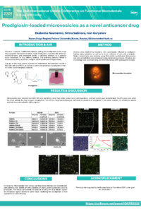
Cancer is a complex, multifactorial disease, making the development of new drugs with a targeted mechanism of action crucial. Prodigiosin, a pigment with antitumor, anti-inflammatory, and antioxidant activity, is attracting attention as a potential active component for drug delivery systems. One promising delivery method is microvesicles (MVs), which can transport active substances to target tissues.
The aim of this study was to produce and characterize microvesicles induced in HEK 293 cells and MSCs, as well as to confirm the presence of prodigiosin in them for further use in therapeutic systems. Methods: Vesicles were obtained by incubation with cytochalasin, followed by prodigiosin loading using sonication, as well as by direct incubation of cells with prodigiosin. Protein concentration was analyzed using BCA protein quantification. Microvesicle size and concentration were measured using nanotracking analysis. Structure and morphology were examined using dark-field microscopy with hyperspectral analysis.
Results: Microvesicles were obtained from HEK 293 cells and MSCs, which had similar protein levels and abundance, and their stability was demonstrated. The MV sizes were within the range suitable for drug transport, ranging from 100-200 nm. Hyperspectral analysis confirmed the presence of prodigosin in the loaded vesicles; the reflectance spectra matched those characteristic of this pigment.
Conclusions: Microvesicles from various cell lines were obtained and characterized, demonstrating their stability and the possibility of loading active substances, such as prodigiosin. The results demonstrate the potential of using MV as a delivery system for therapeutic agents directly to tumor tissue, facilitating the development of new approaches to cancer therapy.
The study was supported by the Russian Science Foundation (RSF) under grant No. 25-25-00011.
Keywords
Prodigiosin
microvesicles
Poster
Poster Naumenko et al sciforum-182261.pdf
Dual-Loaded Curcumin and Phenformin Copolymer Micelles for Targeted Colorectal Cancer Therapy via Mitochondrial Stress and Metabolic Modulation
Supercritical-Assisted Design of SiO₂–TiO₂ Functionalized PLGA Scaffolds for Controlled Drug Delivery Applications