Event submissions
Published
This submission belongs to the session S6. Biomaterials and Implantable Devices for Healthcare of the event The 2nd International Online Conference on Functional Biomaterials
Published date
03 Jul, 2026
Academic Editor
'%3e%3cpath%20d='M12.6657%2010.9104C12.4185%2010.325%2012.0599%209.79327%2011.6097%209.34482C11.1609%208.89508%2010.6293%208.5365%2010.0442%208.28888C10.0389%208.28626%2010.0337%208.28495%2010.0285%208.28233C10.8446%207.69279%2011.3752%206.73249%2011.3752%205.64904C11.3752%203.8542%209.92103%202.39999%208.1262%202.39999C6.33136%202.39999%204.87715%203.8542%204.87715%205.64904C4.87715%206.73249%205.40774%207.69279%206.22393%208.28364C6.21869%208.28626%206.21345%208.28757%206.20821%208.29019C5.62129%208.5378%205.09463%208.89284%204.64265%209.34613C4.1929%209.79493%203.83432%2010.3266%203.58671%2010.9117C3.34345%2011.4845%203.21226%2012.0987%203.20023%2012.7209C3.19988%2012.7349%203.20233%2012.7488%203.20744%2012.7619C3.21255%2012.7749%203.22022%2012.7867%203.22998%2012.7968C3.23975%2012.8068%203.25142%2012.8147%203.26431%2012.8202C3.2772%2012.8256%203.29105%2012.8284%203.30504%2012.8284H4.09109C4.14874%2012.8284%204.19459%2012.7825%204.1959%2012.7262C4.2221%2011.7148%204.62823%2010.7676%205.34617%2010.0497C6.08899%209.30683%207.0755%208.89808%208.1262%208.89808C9.17689%208.89808%2010.1634%209.30683%2010.9062%2010.0497C11.6242%2010.7676%2012.0303%2011.7148%2012.0565%2012.7262C12.0578%2012.7838%2012.1037%2012.8284%2012.1613%2012.8284H12.9474C12.9613%2012.8284%2012.9752%2012.8256%2012.9881%2012.8202C13.001%2012.8147%2013.0126%2012.8068%2013.0224%2012.7968C13.0322%2012.7867%2013.0398%2012.7749%2013.0449%2012.7619C13.0501%2012.7488%2013.0525%2012.7349%2013.0522%2012.7209C13.0391%2012.0947%2012.9094%2011.4855%2012.6657%2010.9104ZM8.1262%207.9024C7.52486%207.9024%206.9589%207.6679%206.53312%207.24211C6.10733%206.81633%205.87283%206.25037%205.87283%205.64904C5.87283%205.0477%206.10733%204.48174%206.53312%204.05596C6.9589%203.63018%207.52486%203.39567%208.1262%203.39567C8.72753%203.39567%209.29349%203.63018%209.71927%204.05596C10.1451%204.48174%2010.3796%205.0477%2010.3796%205.64904C10.3796%206.25037%2010.1451%206.81633%209.71927%207.24211C9.29349%207.6679%208.72753%207.9024%208.1262%207.9024Z'%20fill='%235D1EE1'/%3e%3c/g%3e%3c/svg%3e) Meital Zilberman
Meital ZilbermanCitation
Kesavamoorthy Muthu, Thilanka N. Haththotuwa, Aditya Joshi, George Dias, Mark Staiger, Validation of clinical CT-based volumetric analysis using Ti implants for in vivo monitoring of Mg-based orthopaedic devices, in Proceedings of The 2nd International Online Conference on Functional Biomaterials, 8 July–10 July 2026, MDPI: Basel, Switzerland
Share
Email
Facebook
Twitter
LinkedIn
Validation of clinical CT-based volumetric analysis using Ti implants for in vivo monitoring of Mg-based orthopaedic devices
'%3e%3cpath%20d='M12.6647%2010.9104C12.4176%2010.325%2012.0589%209.7933%2011.6088%209.34485C11.16%208.89511%2010.6283%208.53653%2010.0432%208.28891C10.038%208.28629%2010.0327%208.28498%2010.0275%208.28236C10.8437%207.69282%2011.3743%206.73252%2011.3743%205.64907C11.3743%203.85423%209.92005%202.40002%208.12522%202.40002C6.33038%202.40002%204.87618%203.85423%204.87618%205.64907C4.87618%206.73252%205.40677%207.69282%206.22296%208.28367C6.21772%208.28629%206.21248%208.2876%206.20724%208.29022C5.62031%208.53783%205.09365%208.89287%204.64167%209.34616C4.19192%209.79496%203.83334%2010.3266%203.58573%2010.9117C3.34247%2011.4846%203.21128%2012.0987%203.19925%2012.721C3.1989%2012.735%203.20135%2012.7489%203.20646%2012.7619C3.21157%2012.7749%203.21924%2012.7868%203.22901%2012.7968C3.23877%2012.8068%203.25045%2012.8148%203.26334%2012.8202C3.27623%2012.8256%203.29007%2012.8284%203.30406%2012.8284H4.09012C4.14776%2012.8284%204.19362%2012.7825%204.19493%2012.7262C4.22113%2011.7148%204.62726%2010.7676%205.34519%2010.0497C6.08802%209.30686%207.07452%208.89811%208.12522%208.89811C9.17592%208.89811%2010.1624%209.30686%2010.9052%2010.0497C11.6232%2010.7676%2012.0293%2011.7148%2012.0555%2012.7262C12.0568%2012.7839%2012.1027%2012.8284%2012.1603%2012.8284H12.9464C12.9604%2012.8284%2012.9742%2012.8256%2012.9871%2012.8202C13%2012.8148%2013.0117%2012.8068%2013.0214%2012.7968C13.0312%2012.7868%2013.0389%2012.7749%2013.044%2012.7619C13.0491%2012.7489%2013.0515%2012.735%2013.0512%2012.721C13.0381%2012.0947%2012.9084%2011.4856%2012.6647%2010.9104ZM8.12522%207.90243C7.52388%207.90243%206.95792%207.66793%206.53214%207.24215C6.10636%206.81636%205.87185%206.2504%205.87185%205.64907C5.87185%205.04773%206.10636%204.48177%206.53214%204.05599C6.95792%203.63021%207.52388%203.3957%208.12522%203.3957C8.72655%203.3957%209.29252%203.63021%209.7183%204.05599C10.1441%204.48177%2010.3786%205.04773%2010.3786%205.64907C10.3786%206.2504%2010.1441%206.81636%209.7183%207.24215C9.29252%207.66793%208.72655%207.90243%208.12522%207.90243Z'%20fill='%235D1EE1'/%3e%3c/g%3e%3c/svg%3e)
Kesavamoorthy Muthu 1

Thilanka N. Haththotuwa 2
Aditya Joshi 1
George Dias 2
Mark Staiger 1
1. Department of Mechanical Engineering, University of Canterbury, Christchurch, New Zealand, New Zealand
2. Department of Anatomy, University of Otago, Dunedin, New Zealand, New Zealand
Abstract
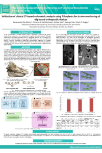
INTRODUCTION: In situ quantification of residual implant volume is desirable in long-term Mg implant studies where repeated ex vivo measurements require euthanising animals. Although clinical X-ray computed tomography (CT) is commonly used for in vivo imaging, its accuracy for measuring implant volumes against CAD models or ex vivo micro-CT is still lacking. The study represents a preliminary methodological validation of CT-based volumetric analysis using Ti implants and CAD reference before application to degrading Mg-based devices.
METHODS: Unstable zygomatic arch fractures were created in three farm pigs and stabilised with craniofacial plate-screw systems. One animal received the Ti-6Al-4V system on the right arch and the ZX10 Mg device on the left, while others received ZX10 devices on both sides. CT scans were acquired at 1, 3, 6, and 12 months, with ex vivo micro-CT performed after one year following euthanasia. Implant segmentation was performed in 3D Slicer using modality-specific intensity thresholds, optimised and applied consistently within each modality. Volumetric analysis of the combined Ti plate-screw system at one year was performed by a single observer.
RESULTS: Ex vivo micro-CT data were highly accurate, with a mean error of 0.944 %, whereas clinical CT showed a higher deviation with a mean error of 9.107 %. The mean clinical CT error may still be acceptable for longitudinal monitoring depending on the magnitude of implant degradation over time. The coefficient of variation was 0.212 % for micro-CT and 1.054 % for clinical CT, indicating good reproducibility for both modalities. The deviation in clinical CT measurements is attributed to partial-volume effects and limited spatial resolution.
CONCLUSIONS: CT-based volumetric analysis of Ti implants demonstrated good reproducibility but lower accuracy than micro-CT. Despite systematic deviation, CT-based analysis is reliable for longitudinal in vivo monitoring when measurement bias is considered. Further studies are underway to assess ZX10 implants which may behave differently due to gradual in vivo degradation altering their volume and geometry.
Keywords
Titanium implants
Magnesium-based implants
Craniofacial fixation
Clinical computed tomography (CT)
Micro-computed tomography (micro-CT)
Volumetric analysis
Poster
IOCFB-Poster-Kesav.pdf
Doxorubicin-loaded Cholesterol-Glycopolymer Nanomicelles with Autophagy Activator Rapamycin for Synergistic Glioblastoma Cell Inhibition
Tantalum-Doped 58S Bioactive Glass Coatings for Corrosion-Resistant CoCrMo Implants