
Cells Webinar | Beyond the Bulk: Decoding Cellular Architecture with Spatial Biology
the chair
27th Cells Webinar
Beyond the Bulk: Decoding Cellular Architecture with Spatial Biology
The life sciences are undergoing a fundamental transition: moving away from the reductive analysis of dissociated tissues toward a "spatial forensics" approach that preserves cellular context. While single-cell sequencing has provided an exhaustive list of molecular players, it remains "context-blind" to the structural logic of the tissue microenvironment.
In this webinar, I will be exploring the technological metamorphosis from classical immunohistochemistry to modern high-plex spatial multiomics. I will move beyond the "cellular smoothie" tissue destruction problem to discuss how researchers are now performing in situ interrogation to uncover biological logic within preserved cellular neighborhoods.
Key learning objectives include:
· Engineering Strategies for Complexity: A critical evaluation of iterative biochemical cycles (seqIF™) versus molecular encoding (DNA barcoding) for multiplexed immunofluorescence.
· Overcoming the "Transcriptomic Trilemma": Strategies to balance spatial resolution, analytical throughput, and transcriptomic depth in experimental design.
· Functional Symbiosis: How to integrate targeted protein imaging with unbiased spatial proteomics to capture the "functional ground truth" of a sample.
· From Discovery to Routine Tool: Addressing the urgent need for cross-platform standardization and the synchronization of multi-omic data layers to improve drug discovery and precision medicine.
· Imaging Issues and Consequent Errors: Addressing quantification problems when Spatial Biology images are not captured appropriately, and when results are shown in reports with comparisons of brightness levels with misleading brightness comparisons, quantitation from these images provides erroneous measurements.
· The Capabilities of the 10x Genomics Atera Platform: The culmination of over a decade of spatial and single-cell innovations, the Atera In Situ platform redefines what's possible with unmatched discovery power, unparalleled throughput and performance, and unrivaled capabilities all at whole transcriptome scale. You will learn (a) how to get the complete picture with whole transcriptome coverage of 18,000+ protein-coding genes and analyze up to 800 cm2 whole transcriptome samples a year, (b) stack up to 1,000 custom genes onto whole transcriptome panels, (c) leverage stackable targeted panels up to 2,000-plex, or (d) create fully custom 2,000-gene panels tailored to researchers’ needs.
Date: 2 December 2026
Time: 17:00 CET | 11:00 EST
Webinar ID: 832 1543 6063
Webinar Secretariat: journal.webinar@mdpi.com
Meet Our Chair
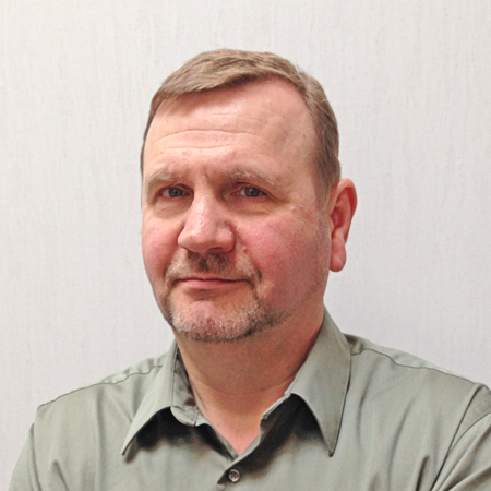
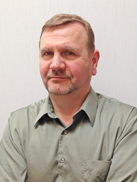
Dental Basic Sciences, University of Minnesota, Minneapolis, USA;Dr. Alexander E. Kalyuzyhny received his B.S. and M.S. from the Department of Biochemistry & Biophysics. He studied the effects of narcotic substances on early morphogenesis and investigated mechanisms underlying opiate-induced antinociception. Since 1998, Dr. Kalyuzhny has been involved with opioid research, teaching, and editorial work. He also served as a manager of the antibody validation and ELISpot laboratory at Bio-Techne, Inc., where he was involved with the development and IHC validation of antibodies for research and clinical applications. His research interests are focused on developing high-sensitivity multi-plex IHC spatial biology techniques to probe the micro-environment of various human and animal tissues. Dr. Kalyuzhny is the editor of 7 books on IHC and ELISpot, the author of an IHC book for non-experts, and has authored 70 papers in peer-reviewed journals and chapters in textbooks.
Meet Our Speakers
Dental Basic Sciences, University of Minnesota, Minneapolis, USA;Dr. Alexander E. Kalyuzyhny received his B.S. and M.S. from the Department of Biochemistry & Biophysics. He studied the effects of narcotic substances on early morphogenesis and investigated mechanisms underlying opiate-induced antinociception. Since 1998, Dr. Kalyuzhny has been involved with opioid research, teaching, and editorial work. He also served as a manager of the antibody validation and ELISpot laboratory at Bio-Techne, Inc., where he was involved with the development and IHC validation of antibodies for research and clinical applications. His research interests are focused on developing high-sensitivity multi-plex IHC spatial biology techniques to probe the micro-environment of various human and animal tissues. Dr. Kalyuzhny is the editor of 7 books on IHC and ELISpot, the author of an IHC book for non-experts, and has authored 70 papers in peer-reviewed journals and chapters in textbooks
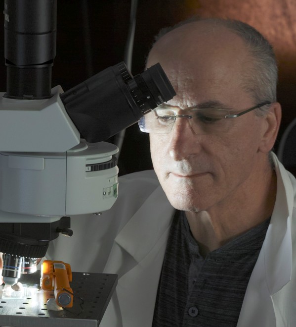
Mr. Jerry Sedgewick
Image Analysis Consulting, Minneapolis, USA;Mr. Gerald (Jerry) Sedgewick is a scientific imaging consultant specializing in computer-aided quantification from microscope images and in image post-processing. Among microscopists and researchers in biology, he pioneered the transition from film-based imaging to digital with books and articles on scientific imaging. He has co-authored papers in Science, PNAS, Circulation and other peer-reviewed publications; conducted workshops on scientific imaging/quantitation at major universities; trained students at Woods Hole; and developed patentable imaging instrumentation for a proteomics scanning device that won the Red Dot Award. His passion is to train scientists to take images correctly for subsequent quantitation.
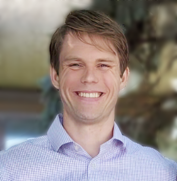
Dr. Daniel Moline
10x Genomics, California, USA;Dr. Daniel Moline received his doctoral training at the University of Chicago, where he worked in the lab of Dr. Donald Vander Griend, pursuing translational research in the prostate cancer space. Since the completion of his graduate degree in late 2020, he has worked at 10x Genomics, first as a Field Application Scientist and now as a Science & Technology advisor. Dr. Moline is a specialist in the field of Spatial Biology applications. He frequently engages with scientists from both academic and industrial backgrounds, offering his expert advice and troubleshooting solutions for a range of experimental setup challenges.
This is a FREE webinar. After registering, you will receive a confirmation email containing information on how to join the webinar. Registration with academic institutional email addresses will be prioritized.
Certificates of attendance will be delivered to those who attend the live webinar.
Unable to attend? Register anyway, and we'll let you know when the recording is available to watch.
Register for free here!
| Speaker/Presentation | Time (CET) | Time (EST) |
| MDPI Introduction | 17:00 - 17:10 | 11:00 - 11:10 |
| Dr. Alexander E. Kalyuzhny Chair Introduction |
17:10 - 17:20 | 11:10 - 11:20 |
| Jerry Sedgewick Imaging Issues and Consequent Errors |
17:20 - 17:40 | 11:20 - 11:40 |
| Dr. Daniel Moline Unlocking Spatial Biology with Atera |
17:40 - 18:00 | 11:40 - 12:00 |
| Dr. Alexander E. Kalyuzhny Mapping Life in Context |
18:00-18:20 | 12:00 - 12:20 |
| Q&A | 18:20 - 18:55 | 12:20 - 12:55 |
| Dr. Alexander E. Kalyuzhny Closing of Webinar |
18:55 - 19:00 | 12:55 -13:00 |
Spatial Biology: Decoding Cellular Complexity in Tissues
Guest Editor: Dr. Alexander E. Kalyuzhny
Deadline for Manuscript Submission: 31 October 2026