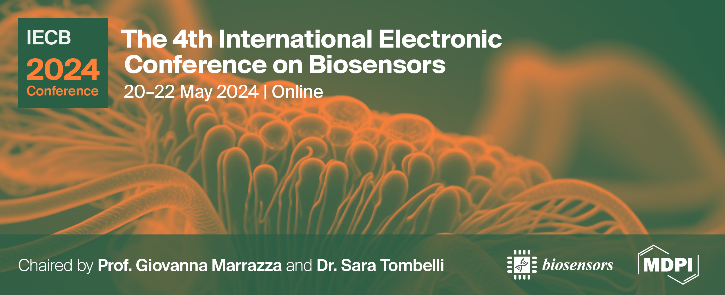
Please login first

List of accepted submissions


Show results per page
Find papers
 
  • Open access
  • 0 Reads
Developing a wearable sensing platform for well-being monitoring in individuals with dopamine-related neurological disorders

Neurological disorders are heterogeneous diseases that affect the body’s autonomic, peripheral and central nervous system. These disorders gradually diminish cognitive and motor abilities, hindering daily activities and resulting in a loss of independence. Detection and continuous monitoring of protein biomarkers involved in these disorders are painful and uncomfortable as they are found in hard-to reach body fluids such as cerebrospinal fluid or blood. However, several biomarkers or metabolites reside in peripheral biofluids such as saliva, urine and sweat, facilitating the development of wearable biosensors for their non-invasive detection.

Electrochemical sensors offer amazing potential for the development of wearable devices because they can be miniaturized and integrated into comfortable wearable devices (bracelets, smartwatches, etc.).

This study introduces a new analytical wearable platform based on screen-printed electrodes for monitoring dopamine in real samples by differential pulse voltammetry. The sensor was created by electrodepositing a biocompatible polymeric conductive layer onto carbon screen-printed electrodes. The polymeric layer was further modified by electrodepositing gold nanoparticles, in order to direct detect electroactive molecules. The modified platforms were characterized using cyclic voltammetry and electrochemical impedance spectroscopy. The experimental conditions for dopamine detection in body fluids were studied and optimized. Linear calibration curve in the range of 0–100 μM with a limit of detection of 0.002 μM for dopamine determination was obtained. The analytical performances of the sensors in terms of reproducibility and selectivity were also evaluated.

Acknowledgements

This work was supported by the European Union by the NextGenerationEu project ECS00000017 ‘Ecosistema dell’Innovatione’ Tuscany Health Ecosystem (THE, PNRR, Spoke 3: Nanotechnologies for diagnosis and therapy).

  • Open access
  • 0 Reads
Monitoring salivary biomarkers in astronauts during spaceflight: a versatile and reusable portable microfluidic biosensor based on functionalized superparamagnetic microbeadsfor performing multiplex chemiluminescent immunoassays

Introduction. Portable and simple analytical devices to be used for routinary self-diagnostic applications are particularly attractive for space applications since the crewmembers must live in a microgravity environment for several months. The proposed work in the project APHRODITE (financed by Italian Space Agency) focused on the optimization of a dual-chemiluminescent immunoassay implemented onto a portable and easy-to-use platform for the quantification of two salivary hormones, cortisol and dehydroepiandrosterone (DHEA).

Method. The method is based on the use of immunological techniques combined with chemiluminescent (CL) detection by exploiting a microfluidic channel integrated with a dedicated detector. Two aliquots of magnetic beads functionalized with anti-cortisol and anti-DHEA antibodies, respectively, were entrapped by magnets in two different areas along the microchannel. A solution containing the sample and the peroxidase conjugates of both DHEA and cortisol was injected to enable competition for binding the antibodies immobilized on MB surfaces. By adding the proper CL substrate, it was possible to monitor the CL signal in real time by employing an array of hydrogenated amorphous silicon (a-Si:H) photosensors. Once the assay was completed, by removing the magnets and performing the washing step, the microchannel was clean and ready to run a new assay.

Results. With this system, taking advantage of the possibility of magnetically confining the immunoprobes, chemiluminescence detectability, and photosensor sensitivity, the accurate quantification of target analytes down to 0.1 ng mL-1 for cortisol and 0.05 ng mL-1 for DHEA were obtained with high specificity and multiplexing ability.

Conclusion. Results confirmed the good detection capabilities and assay applicability of the proposed system, prompting the development of innovative universal tools used for multiplex assays that allow a health-related panel of biomarkers to be simultaneously monitored through a single analysis.

  • Open access
  • 0 Reads
Development of a Chemiluminescent immunoassay for the detection of Dehydroepiandrosterone in counterfeit cosmetic products

Introduction. Many countries have developed regulatory lists of banned or restricted cosmetic ingredients for ensuring the safety of cosmetic products. Among the banned substances, dehydroepiandrosterone (DHEA) is an hormone which can affect the biological processes in humans even at a very low level, but it is often illegally added to cosmetic formulations since it has an anti-ageing effect. The aim of this study is to develop a chemiluminescent (CL)-based immunoassay for the detection and quantification of DHEA in cosmetics.

Method. The method involved the development of an integrated lab-on-a-chip platform, the assay was carried out in solution, and the detection was performed inside microwells coupled to a dedicated CL detector.

In this approach, magnetic beads (MBs), used as platform for the competitive immunoassay, were functionalized with anti-DHEA antibodies and were incubated with a solution containing the sample and the peroxidase conjugates of DHEA to provide competition for binding the antibodies. After the washing step, the MBs were added into a black microwell plate with a transparent bottom which was aligned with an array of hydrogenated amorphous silicon photosensors. After the addition of the proper CL substrate, it was possible to monitor the CL signal in real time.

Results. The developed method allowed for the accurate quantification of DHEA down to 20 ppb, and tests were performed on spiked cosmetic samples and suitably pre-treated. 

Conclusion. The proposed assay demonstrated the possibility to exploit a rapid and simple CL-immunoassay for detecting counterfeit at very low level in complex cosmetic matrices. The method can be optimized in the future for different kinds of banned ingredients, allowing the number of target analytes that can be detect into a single analysis to be increased by exercising a multiplexing approach.

  • Open access
  • 0 Reads
Design of Internet of Things-enabled textile-based biosensors

During a health crisis or pandemic, people with breathing issues may find it challenging to receive timely medical attention as access to healthcare services is limited. Breathing difficulties can contribute to anxiety, stress, and uncertainty, and fear associated with a health crisis can further negatively impact mental health. To effectively tackle this situation, it is essential to diagnose these issues early. Furthermore, the primary symptom of such diseases is difficulty in breathing, accompanied by coughing. To identify these conditions, various sensor technologies such as plethysmography sensors, pressure transducers, acoustic sensors, accelerometers, gyroscopes, etc., were used. However, the use of these sensors poses certain challenges in terms of accuracy, calibration issues, discomfort, influence of body position, noise interference, cost, reliability in dynamic environments, and user acceptance. In this work, a textile-based sensor using conductive thread is designed and developed for the measurement of respiration rate. Further, the conductive thread is combined with woollen material to form a chest band that can be affixed to any person. Also, the developed sensor in the form of a chest band is integrated into the Internet of Things (IoT) controller, which measures the respiratory rate of the users while they perform various activities such as walking, running, practicing exercises, standing still, etc. Results demonstrate that the developed sensor can store the respiratory rate in the IoT cloud platform. Further, it is observed that the frequency of the acquired signals due to respiration changes for various activities. Additionally, the increase in frequency increases the noise in the acquired signal, and it is removed using a filter algorithm. This work appears to be of high clinical significance since the developed sensor diagnoses the breathing disorders in terms of respiration rate.

  • Open access
  • 0 Reads
Machine learning-driven calibration in impedimetric biosensors

Electrochemical biosensors are leading the way in the development of point-of-care devices. Many of these biosensors predominantly use electrochemical impedance spectroscopy for their detection mechanism. The standard method for creating calibration curves in impedimetric biosensors is to fit the impedance spectrum to an equivalent circuit model(ECM). The calibration curve is then created using the circuit parameter that shows the maximum variance. This standard approach has multiple limitations. First, in a complicated system, the identification of an ECM that correctly describes the system is not a trivial task as it might involve complicated processes. Second, in many cases, multiple ECMs could fit a given impedance spectrum which could introduce errors or biases in the analysis. To overcome these limitations, in this work, we use machine learning (ML) algorithms to develop calibration curves for impedimetric sensors without the use of equivalent circuit models or by manually picking points from the impedance spectrum. For this, raw impedance spectrum corresponding to different impedimetric sensors were obtained from the literature and a labeled dataset was created for training the ML model. Principal component analysis was done to extract the features that show maximum variance. These features were then used to train multiple machine-learning models to create a calibration curve. The errors of the different models are compared to identify the best-performing ML model. Finally, a comparison was made between the calibration curve obtained from the conventional approach and that obtained from the ML model. This comparison shows that errors were lower for predictions made using the calibration curve obtained from the ML model.

  • Open access
  • 0 Reads
An innovative vapor-phase synthesis approach to obtain MIP-based optical sensors for label free detection of quercetin

Introduction
Quercetin (QU) stands out as the most abundant dietary bioflavonoid supplement, renowned for its
anti-inflammatory, anti-oxidative, and anti-carcinogenic properties. However, the potential for harmful
effects with excessive use necessitates vigilant monitoring. Consequently, continuous screening of
QU assumes paramount importance 1 . Various analytical techniques are employed for QU detection
but simple and rapid approach are needed 1 . Here, we proposed the use of an innovative vapor-phase
polymerization approach to obtain polypyrrole (PPy)-based molecularly imprinted polymers (MIPs) for
QU, within the nanostructure of porous silicon (PSi) photonic crystals, used as interferometers. The
aim is to combine the recognition abilities of the MIP receptors with the optical properties of PSi to
obtain a robust, selective and reusable sensor for QU.
Materials and methods
Porous silicon (PSi) photonic crystals were initially fabricated through an electrochemical etching
procedure on p++ silicon wafers. Subsequently, PSiO 2 layers were obtained via thermal oxidation of
PSi at 1000°C for 10 minutes. In a subsequent step, the QU target was anchored within the PSiO 2
layer, employing an imidazolide-mediated coupling reaction. Afterwards, a vapor-phase
polymerization of Py, developed by our group 2 ,was performed to obtain a thin film around the target.
Successive removal of QU molecules from the polymer matrix produced the imprinted cavities and
then the MIP.
Results and conclusions
The functionalization steps were monitored by UV-VIS spectroscopy, which demonstrated the
effectiveness of the MIP synthesis on PSiO 2 . Preliminary detection tests showed that the sensor can
detect QU in aqueous solutions in a dynamic concentration range between 0.005 to 0.1 mM.
Moreover, the sensor selectivity was tested upon exposure to other antioxidant agents such as gallic
acid and vanillic acid recording in each case a higher sensor response for the target molecule.
Repeatability and stability tests along with QU detection tests in real matrices are in progress.
References
1. doi:10.1080/10408347.2023.2269421.
2. doi:10.1002/SMLL.202302274.

  • Open access
  • 0 Reads

An innovative and versatile reconfigurable sensor for the detection of biomolecules via metal ion-mediated recognition

Introduction

Reconfiguration of chemical sensors, meaning the capacity to adapt the sensor to new target analytes, is potentially game changing and would enable rapid and cost-effective reactions to dynamic changes occurring at different levels, although this is still a challenge. Here, we report on a reconfigurable label-free optical sensor leveraging versatile immobilization of a metal ion chelating agent on a nanostructured porous silica (PSiO2) optical transducer for the detection of different biomolecules.

Method

PSiO2 scaffolds were functionalized with a modified silane, GLYMO-IDA, preliminarily derived from a condensation reaction between 3-glycidoxypropyltrimethoxysilane (GLYMO) and iminodiacetic acid (IDA). When exposed to aqueous solutions of metal ions (e.g., Zn2+, Ni2+, Cu2+, Fe3+), GLYMO-IDA exhibits the ability to chelate them. The as-modified silane was employed as a receptor for biomolecules, leveraging the metal ions as pivotal points. The metal ion on GLYMO-IDA was easily switched by treating the PSiO2 samples with a complexing agent.

Results

Successful functionalization of PSiO2 scaffolds with the artificial receptors for various biomolecules was corroborated via UV-VIS spectroscopy and XPS analysis. After switching the metal ions from Cu2+ to Zn2+, the sensor was used in carnosine detection tests, proving its ability, in both configurations, to detect the target in a concentration range between 0.1 and 1 mM. Alternatively, by switching Cu2+ with Fe3+ ions, the sensor was successfully used for adenosine triphosphate (ATP) detection. A satisfactory sample-to-sample reproducibility was obtained (RSD: ~15%), along with an excellent repeatability (RSD: 2.5%) and stability (30 days).

Conclusions

A reconfigurable sensing platform was developed, and sensor reconfiguration was achieved by switching the metal ions from Cu2+ to Zn2+ and testing its ability to detect the dipeptide carnosine. Additionally, by switching Cu2+ with Fe3+ ions, the sensor was able to detect the target ATP, demonstrating effective reconfiguration of the sensor with the proposed surface chemistry.

  • Open access
  • 0 Reads
Development of an implantable sensor for recording neural activity in the auditory pathway of rats

Introduction: Ensuring the secure and efficient application of invasive microelectrode arrays (MEAs) in chronic experiments is crucial. Therefore, this study aims to develop an implantable multi-channel MEA for simultaneous neural activity recording in the auditory cortex (AuC) and medial geniculate nucleus (MGN) of rats. Methods: We utilized 32-channel tungsten tetrode arrays, comprising eight tetrodes, each constructed from four twisted microwires with a diameter of 15μm, coated with 1.5μm-thick isonel insulation. The tetrode lengths varied based on the region (5.4 or 5.8mm), maintaining a minimum spacing of 300μm. The tetrodes were threaded through an acrylic template (5 x 4mm) containing stereotaxic coordinates of target regions in the left hemisphere. Subsequently, the wires were secured with composite resin, and a printed circuit board connected to an analog headstage (Plexon) was affixed to the template. Array coordinates were organized based on a rat brain atlas, focusing on auditory regions of interest: primary AuC (Au1, tetrodes 1Au1 and 2Au1), secondary AuC dorsal (AuD) and ventral (AuV), MGN ventral (1MGV and 2MGV) and dorsal (1MGD and 2MGD). Results: Following implantation surgery, extracellular recordings and triaxial inertial sensor data were collected from behaving Wistar rats (n=5). Results indicate that tetrodes in Au1 regions (1Au1: -0.483 ± 0.008, 2Au1: -0.618 ± 0.008), AuV (-0.618 ± 0.005), and AuD (-0.472 ± 0.007) exhibited signals with greater amplitudes compared to tetrodes in MGV (1MGV: -0.228 ± 0.006, 2MGV: -0.256 ± 0.004) and MGD (1MGD: -0.193 ± 0.008, 2MGD: -0.162 ± 0.004). Conclusions: These findings suggest that the developed tetrode array sensor may function as an electrophysiologically stable device for recording neural activity in the AuC and MGN of rats.

  • Open access
  • 0 Reads
AI-Driven Improvements in Electrochemical Biosensors for Effective Pathogen Detection at Point-of-Care

The rapid and accurate detection of pathogens is vital for effective disease management and control. This paper introduces a novel approach to pathogen detection by integrating artificial intelligence (AI) into electrochemical biosensors. Real-world samples can present background interference from other analytes and unwanted noise in the signal, particularly when utilizing portable point-of-care devices. To overcome these challenges, we propose an intelligent electrochemical device optimized for improved performance in detecting viral pathogens. Our approach involves two key AI strategies. First, a denoising autoencoder is employed to effectively remove noise from the electrochemical signals, bringing the performance of portable devices on par with their standalone counterparts. This enhancement is crucial for point-of-care applications where environmental and operational factors often compromise data quality. Second, we utilize an Artificial Neural Network (ANN) to detect the presence of background interference. Smartphones are often used as interface for portable electrochemical devices, our approach leverages the computational capabilities of smartphones to run the AI algorithms for processing the electrochemical signals in real-time. The proposed system has been validated using COVID-19 data, demonstrating its potential as a powerful tool in the rapid and accurate detection of SARS-CoV-2 and other pathogens. The integration of AI into electrochemical biosensing offers a more reliable and accessible option for healthcare professionals and researchers.

  • Open access
  • 0 Reads
DEVELOPMENT OF A FLEXIBLE PIEZOELECTRIC BIOSENSOR THAT INTEGRATES BATiO3–POLY(DIMETHYLSILOXANE) FOR POSTURE CORRECTION APPLICATIONS

The global issue of prolonged desk work, which leads to poor posture, has prompted a growing need for effective solutions. This study explores a transformative approach developing and creating a flexible Barium titanate–poly(dimethylsiloxane) (BaTiO3–PDMS) piezoelectric biosensor tailored to addressing posture challenges1. Harnessing the distinctive properties of BaTiO3, this innovative biosensor has the potential to revolutionize posture correction strategies. First, piezoelectric BaTiO3 nanoparticles (NPs) were synthesized using a sol-gel method, utilizing barium acetate and titanium tetrabutanolate as precursors. The characterization of these NPs was conducted using XRD and TEM. A BaTiO3–PDMS composite was then formed by combining PDMS polymer and its cross-linking agent in a 10:1 weight ratio, with the addition of 40 grams of BaTiO3 nanoparticles.

The NPs were manually dispersed in the polymer mixture for 5 minutes. The mixture was cured at 100°C for 1 hour. Evaporation was carried out to achieve a thickness of 2.572 kÅ on both surfaces. Copper was evaporated in order to obtain a conductive material on the surface of the BaTiO3–PDMS. The outcome was a 0.7 mm wide BaTiO3–PDMS biosensor, as measured with a digital multimeter. In testing, the BaTiO3–PDMS sensor generated a 400 mV signal on the oscilloscope, indicating its potential effectiveness for posture correction applications (Fig.1). In conclusion, our exploration of the BaTiO3–PDMS flexible piezoelectric biosensor for posture correction unveils promising initial results. This research is a vital step toward attaining a thorough grasp of this biosensor's capabilities. Future research and optimization are necessity to unlock the optimum amount of BaTiO3–PDMS to use in addressing the ongoing challenge of slumped posture.

Reference:

[1] Jeronimo K, Koutsos V, Cheung R, Mastropaolo E. PDMS-ZnO Piezoelectric Nanocomposites for Pressure Sensors. Sensors. 2021; 21, 5873.

Top