Cell-to-Cell Metabolic Cross-Talk in Physiology and Pathology
Part of the International Online Conference on Cells series
17 December 2020–17 January 2021
exosomes, cell death, cell metabolism, diseases, Autophagy, embriomorphogenesis, Cell Migration, Cell Signaling
- Go to the Sessions
-
- A. Cell Cycle Regulators: the Cross-talk with Metabolism
- B. Exosomes and Extracellular Vesicles in Health and Disease
- C. The Crosstalk Between Cell Adhesion and Metabolism
- D. The cross talk between cell death regulation and metabolism
- E. Compartmentilisation of Cellular Signaling
- F. Signaling and Metabolic Cross-talk in the Tumor Microenvironment
- G. The cross-talk between immune cells and tissue microenvironment
- P. Posters
- Event Details
-
- The 'Cell-to-Cell Metabolic Cross-Talk in Physiology and Pathology'-Introduction from Chair
- Welcome from the Chair
- Conference Chairs
- Event Calls
- Sessions
- Cells2020 Live Sessions Programs
- Related Webinars
- Cells2020 Recording
- Instructions for Authors
- List of Accepted Submissions
- Event Awards
- Sponsors and Partners
- Events in series CELLS-2
We are pleased to announce that the Cells2020 is closed!
Thank you for your participation.
The 'Cell-to-Cell Metabolic Cross-Talk in Physiology and Pathology'-Introduction from Chair
Prof. Dr. Ciro Isidoro
Welcome from the Chair
Dear Colleagues,
On behalf of the Scientific Advisory Committee, I am pleased to welcome you to the first electronic conference, Cells 2020, dedicated to the “mechanisms and pathophysiological role of the metabolic cross-talk between the cells”.
Cells 2020 is an online event that covers all aspects of cell metabolic cross-talk.
The event is hosted on Sciforum, the platform developed by MDPI for organizing electronic conferences and discussion groups, and is sponsored by MDPI and the Cells open access Journal.
The theme of the electronic conference is “Cell-to-Cell Metabolic Cross-Talk in Physiology and Pathology” .
The conference will be held from 17 December 2020 to 17 January 2021 on sciforum.net, a platform service for hosting international electronic conferences for scientific communities.
Topical sessions include:
- Cell Cycle Regulators: The Cross-Talk with Metabolism
- Exosomes and Extracellular Vesicles in Health and Disease
- The Cross-Talk between Cell Adhesion and Metabolism
- The Cross-Talk between Cell Death Regulation and Metabolism (Two webinar: December 17)
- The Compartmentalisation of Cellular Signaling
- Signaling and Metabolic Cross-talk in the Tumor Microenvironment (Two webinars: December 18)
- The Cross-Talk between Immune Cells and Tissue Microenvironment
Cells 2020 will allow scientists from all over the world to present their latest research and receive direct feedback from peers, without costs of travel. Cells 2020 will be sponsored by MDPI, Switzerland.
Please note that registration and publishing abstracts/presentations/proceedings are free of charge. Full papers will be selected for publication in a Special Issue of Cells (IF 4.366; with 20% discount on article processing charges): Selected Papers from the International Electronic Conference on Cell-to-Cell Metabolic Cross-Talk in Physiology and Pathology.
We hope that you will join the conference to make use of the presence of internationally renowned scholars to exchange ideas and to merge different areas of expertise into successful collaborations.
Ciro Isidoro, Chairman of the Scientific Advisory Committee

Department of Health Sciences
Università del Piemonte Orientale
Novara (Italy)
Conference Secretariat
Ms. Christina Zhang
M.Sc. Fancy Zhai
email: cells2020@mdpi.com
Follow the conversion on Twitter with @Cells_MDPI
Conference Chairs

Department of Health Sciences, University of Piemonte Orientale, Amedeo Avogadro, Novara, Italy
Qualified FULL PROFESSOR of -Clinical Biochemistry and Clinical Molecular Biology; -Medical Oncology; -General Pathology and Clinical Pathology.
ciro.isidoro@med.uniupo.it
Conference Committee

Institute of Regenerative Medicine and Biotherapy, INSERM, U1183, Hôpital Saint-Eloi, Montpellier, France
christian.jorgensen@inserm.fr

Stephenson Cancer Center, The University of Oklahoma Health Sciences Center, OKC (USA)
danny-dhanasekaran@ouhsc.edu

Department of Pathology and Laboratory Medicine, Weill Cornell Medicine, New York, NY, USA
mtd4001@med.cornell.edu

Department of Pathology and Cell Biology at CUMC
Department of Otolaryngology/Head & Neck Surgery
Herbert Irving Comprehensive Cancer Center
Columbia University Medical Center, NY, USA
gs2157@cumc.columbia.edu

Institute of Research for Food Safety & Health IRC-FSH, University Magna Graecia, Catanzaro, Italy
mollace@unicz.it

Department of Pharmacy, Health and Nutritional Sciences University of Calabria, Rende (Cs) Italy
rosamaria.lappano@unical.it

Associate Professor of Experimental Medicine and Pathophysiology, Dept. of Health Sciences, Università degli Studi di Milano, Milano, Italy
raffaella.chiaramonte@unimi.it

University of Pennsylvania, Children's Hospital of Philadelphia, 1211B Abramson Research Center, 3615 Civic Center Blvd, Philadelphia, PA, USA
bailisw@email.chop.edu

Ph.D. Professor, Laboratory of Molecular Virology, George Mason University Discovery Hall, Manassas, VA, USA
fkashanc@gmu.edu
Call for Submissions
The Cell-to-Cell Metabolic Cross-Talk in Physiology and Pathology (Cells 2020) will be held online from 17 December 2020 to 17 January 2021. This event enables the researchers of cells sciences to present their research and exchange ideas with their colleagues without the need to travel. All proceedings will be published on the conference homepage in an open access format.
Through this event, we aim to cover the following topics:
- Cell Cycle Regulators: the Cross-talk with Metabolism
- Exosomes and Extracellular Vesicles in Health and Disease
- The Crosstalk Between Cell Adhesion and Metabolism
- The Crosstalk between Cell Death Regulation and Metabolism
- The Compartmentilisation of Cellular Signaling
- Signaling and Metabolic Cross-talk in the Tumor Microenvironment
- The Crosstalk between Immune Cells and Tissue Microenvironment
The conference will be completely free of charge—both to attend and for scholars to upload and present their latest work on the conference platform. There will also be the possibility to submit selected papers to the journal Cells (ISSN 2073-4409; CODEN: CELLC6; Impact Factor: 4.366 (JCR 2019) with a 20% discount on the APCs; Cells 2020 offers you the opportunity to participate in this international, scholarly conference without having the concern or expenditure of travel—all you need is your computer and access to the Internet. We would like to invite you to “attend” this conference and present your latest work.
Abstracts (in English) should be submitted by 1 December 2020 online at https://www.sciforum.net/login. For accepted abstracts, the full paper can be submitted by 13 December 2020. The conference itself will be held from 17 December 2020 to 17 January 2021.
We hope you will be able to join this exciting event and support us in making it a success. Cells 2020 is organized and sponsored by MDPI, a scholarly open access publisher based in Basel, Switzerland.
Paper Submission Guidelines
For information about the submission procedure and the preparation of a full presentation, please refer to the "Instructions for Authors".
Critical Dates
Sessions
B. Exosomes and Extracellular Vesicles in Health and Disease
C. The Crosstalk Between Cell Adhesion and Metabolism
D. The cross talk between cell death regulation and metabolism
E. Compartmentilisation of Cellular Signaling
F. Signaling and Metabolic Cross-talk in the Tumor Microenvironment
G. The cross-talk between immune cells and tissue microenvironment
P. Posters
Cells2020 Live Sessions Programs
17 December
| Professor Danny N. Dhanasekaran | Bidirectional Metabolic Signaling loop in Ovarian Cancer Microenvironment |
5:00-5:30 pm |
| Dr. Gloria H. Su | Cellular Complexity of Pancreatic Cancer |
5:30-6:00 pm |
18 December
| Dr. Rosamaria Lappano | Hypoxia-IL1β Signaling Prompts Stimulatory Effects in Both Breast Cancer Cells and Cancer-Associated Fibroblasts |
9:00-9:30 am |
| Professor Dr. Ciro Isidoro | Dysregulation of Glucose Metabolism in Cancer Cells: Role of the Inflammatory Tumor Microenvironment |
10:00-10:30 am |
Related Webinars
| Speaker | Presentation topic | Date and Time (CET) | Registration |
| Dr. Jan Lotvall | Extracellular vesicles as communicators between cells – importance in health and disease |
17 November 9:20-9:50 am |
|
|
Dr. Samir El Andaloussi |
Engineering of extracellular vesicles |
17 November 10:00-10:30 am |
|
| Dr. Davide Medica | Extracellular vesicles derived by human endothelial progenitor cells protect human renal glomerular endothelial cells and podocytes from tumor necrosis factor-alpha injury |
18 November 11:00-11:40 am |
|
| Professor Maria T. Diaz-Meco | Metabolic vulnerabilities in the control of lineage plasticity in cancer |
18 November 3:20-4:00 pm |
|
| Dr. Raffaella Chiaramonte | Extracellular vesicles: new players in multiple myeloma bone marrow reprogramming |
18 November 4:00-4:40 pm |
|
| Dr. Sarah Hurtado-Bagès | The histone variant macroH2A1.1, a master regulator of myogenic cell fusion, adhesion and metabolism |
19 November 10:30-11:00 am |
|
| Prof. Vincenzo Mollace | The role of translocator protein TSPO and intracellular NT-metalloproteinase-2 in dysfunctional myocardial cells in diabetics. |
23 November 9:00-9:30am |
|
| Dr. Lorena Urbanelli | Extracellular vesicles: from endosomal/lysosomal system to extracellular signalling |
23 November 9:40-10:10 am |
|
| Dr. Ravi P. Sahu | 1. Lipid signaling pathway in targeted therapy-mediated microvesicle particles release in lung cancer 2.Exploring the lipid signaling pathway and microRNA-149 in modulating the growth and cytotoxic responses of targeted therapy against lung cancer |
23 November 11:20am -12:00 pm |
|
| Dr. Will Bailis | Regulation of T cells by NAD compartmentalization |
23 November 2:15-2:55 pm |
|
| Professor Fatah Kashanchi | Use of exosomes to communicate between infected and uninfected cells |
25 November 11:30 am-12:00 pm |
|
| Professor Annalisa Chiocchetti | Osteopontin shapes the metastatic niche by engaging ICOS-L |
25 November 9:40-10:10 am |
|
| Professor Antonio Sica | Connections between emergency hematopoiesis and tumor microenvironment |
25 November 10:20-10:50 am |
Cells2020 Recording
In this section, you will find the recordings of the webinars to watch, re-watch and share with your colleagues!
17 November 2020
18 November 2020
19 November 2020
25 November 2020
17 December 2020
18 December 2020
Instructions for Authors
Submissions should be done by the authors online by registering with www.sciforum.net, and using the "Start New Submission" function once logged into system.
- Scholars interested in participating with the conference can submit their abstract (about 150-300 words covering the areas of manuscripts for the proceedings issue) online on this website until 1 December 2020.
- The Conference Committee will pre-evaluate, based on the submitted abstract, whether a contribution from the authors of the abstract will be welcome in Cells 2020. All authors will be notified by 3 December 2020 about the acceptance of their abstract.
- If the abstract is accepted for this conference, the author is asked to submit his/her manuscript, optionally along with a PowerPoint (only PDF) and/or video presentation of his/her paper, until the submission deadline of 13 December 2020.
- The manuscripts and presentations will be available on the Cells 2020 homepage for discussion and rating during the time of the conference 17 December 2020 to 17 January 2021.
- Accepted papers will be published in the proceedings of the conference and journal Cells will publish the proceedings of the conference as a Special Issue. After the conference, the authors are recommended to submit an extended version of the proceeding papers to the Cells Special Issue with a 20% discount on the Article Processing Charges.
Manuscripts for the proceedings issue must have the following organization:
- Title
- Full author names
- Affiliations (including full postal address) and authors' e-mail addresses
- Abstract
- Keywords
- Introduction
- Methods
- Results and Discussion
- Conclusions
- (Acknowledgements)
- References
Manuscripts should be prepared in MS Word and should be converted to the PDF format before submission. The publication format will be PDF. There is no page limit on the length, although authors are asked to keep their papers as concise as possible.
Authors must use the Microsoft Word template to prepare their manuscript. Using the template file will substantially shorten the time to complete copy-editing and publication of accepted manuscripts. Manuscript prepared in MS Word must be converted into a single file before submission. Please do not insert any graphics (schemes, figures, etc.) into a movable frame which can superimpose the text and make the layout very difficult.
- Paper Format: A4 paper format, the printing area is 17.5 cm x 26.2 cm. The margins should be 1.75 cm on each side of the paper (top, bottom, left, and right sides).
- Formatting / Style: Papers should be prepared following the style of Cells 2020 template. The full titles and the cited papers must be given. Reference numbers should be placed in square brackets [ ], and placed before the punctuation; for example [4] or [1-3], and all the references should be listed separately and as the last section at the end of the manuscript.
- Authors List and Affiliation Format: Authors' full first and last names must be given. Abbreviated middle name can be added. For papers written by various contributors a corresponding author must be designated. The PubMed/MEDLINE format is used for affiliations: complete street address information including city, zip code, state/province, country, and email address should be added. All authors who contributed significantly to the manuscript (including writing a section) should be listed on the first page of the manuscript, below the title of the article. Other parties, who provided only minor contributions, should be listed under Acknowledgments only. A minor contribution might be a discussion with the author, reading through the draft of the manuscript, or performing English corrections.
- Figures, Schemes and Tables: Authors are encouraged to prepare figures and schemes in color. Full color graphics will be published free of charge. Figure and schemes must be numbered (Figure 1, Scheme I, Figure 2, Scheme II, etc.) and an explanatory title must be added. Tables should be inserted into the main text, and numbers and titles for all tables supplied. All table columns should have an explanatory heading. Please supply legends for all figures, schemes and tables. The legends should be prepared as a separate paragraph of the main text and placed in the main text before a table, a figure or a scheme.
For further enquiries please contact the Conference Secretariat.
Authors are encouraged to prepare a presentation in PowerPoint, to be displayed online along with the Manuscript. Slides, if available, will be displayed directly in the website using Sciforum.net's proprietary slides viewer. Slides can be prepared in exactly the same way as for any traditional conference where research results can be presented. Slides should be converted to the PDF format before submission so that our process can easily and automatically convert them for online displaying.
Authors are also encouraged to submit video presentations. The video should be no longer than 10 minutes and be prepared with the following formats,
- .MOV
- .MPEG4
- .MP4
- .AVI
- .WMV
- .MPEGPS
- .FLV
The video should be submitted via email before 13 December 2020.
Posters will be available on this conference website during and after the event. Like papers presented on the conference, participants will be able to ask questions and make comments about the posters. Posters that are submitted without paper will not be included in the proceedings of the conference.
It is the authors' responsibility to identify and declare any personal circumstances or interests that may be perceived as inappropriately influencing the representation or interpretation of clinical research. If there is no conflict, please state here "The authors declare no conflict of interest." This should be conveyed in a separate "Conflict of Interest" statement preceding the "Acknowledgments" and "References" sections at the end of the manuscript. Financial support for the study must be fully disclosed under "Acknowledgments" section. It is the authors' responsibility to identify and declare any personal circumstances or interests that may be perceived as inappropriately influencing the representation or interpretation of clinical research. If there is no conflict, please state here "The authors declare no conflict of interest." This should be conveyed in a separate "Conflict of Interest" statement preceding the "Acknowledgments" and "References" sections at the end of the manuscript. Financial support for the study must be fully disclosed under "Acknowledgments" section.
MDPI, the publisher of the Sciforum.net platform, is an open access publisher. We believe that authors should retain the copyright to their scholarly works. Hence, by submitting a Communication paper to this conference, you retain the copyright of your paper, but you grant MDPI the non-exclusive right to publish this paper online on the Sciforum.net platform. This means you can easily submit your paper to any scientific journal at a later stage and transfer the copyright to its publisher (if required by that publisher).
List of accepted submissions (7)
| Id | Title | Authors | Presentation Video | Poster PDF | |||||||||||||||||||||||||||||||||||||
|---|---|---|---|---|---|---|---|---|---|---|---|---|---|---|---|---|---|---|---|---|---|---|---|---|---|---|---|---|---|---|---|---|---|---|---|---|---|---|---|---|---|
| sciforum-034883 | The actin cytoskeleton is a key element of the apoptosome assembly in the developing brain | , , | N/A | N/A |
Show Abstract |
||||||||||||||||||||||||||||||||||||
|
BACKGROUND: The formation of apoptosomes is well-established in the mechanism of programmed cell death. The interaction of APAF-1, caspase-3 and -9, which by adding of cytochrome C only in the presence of macroergs (dATP or ATP) generates apoptosome. Besides the cytoplasmic protein concentration is critical for the assembly of apoptosomes since in vitro it can induced only at values starting from ~2 mg/ml. The study of this feature led us to the discovery of at least one more protein that is critically involved in their formation. METHODS: Cytoplasmic fraction from brain homogenates of newborn rats was obtained by centrifugation. Nucleoside di- and triphosphates, Na+, K+, RNAse A, DNAse1, phalloidin etc., as well as non-muscle F (filamentous) and G (monomeric) actin were added to study their effect on formation of active apoptosomes in the cytoplasm. Preliminary analysis showed that the proteasome activity of the "hydrolyzing peptidyl-glutamyl peptide" constitutes a significant part of all activities in the cleavage of caspase substrates. In this regard, experiments to study the activity of caspases were carried out in the presence of proteasome inhibitors (bortezomib or AdaAhx3L3VS), which do not affect the assembly of apoptosomes. Caspase activity was confirmed by the use of a selective caspase inhibitor emricasane. RESULTS: It was found that manipulations with the cytoplasm (its concentration variation, the presence of monovalent cations, membrane fragments) in a dose-dependent manner nonlinearly led to an increasing of formed apoptosomes and the activity of caspase-3. These modifications changed the protease activity: maximal velocity from 0.15 to 2.4 mkmol/mg/min and Km from 4.2 to 0.13 mkM. A more detailed analysis showed that substances influencing on the assembly and disassembly of actin filaments directly affect both the formation of apoptosomes and the caspase-3 activity. This influence is critically significant, changing the activity by at least an order of magnitude. In this case, the effect of proteins that directly inhibit the activities of caspases (for example, XIAP) did not change. CONCLUSIONS: Thus the actin G/F ratio (balance assembly/disruption of actin filaments) is a key regulator of the assembly of apoptosomes and it depends on the presence of nucleoside triphosphates. It explains the critical dependence of apoptosis on the production of macroergs. |
|||||||||||||||||||||||||||||||||||||||||
| sciforum-034829 | Apoptosomes and proteasomes from exosomes generated by human hematopoietic stem cells | , , | N/A | N/A |
Show Abstract |
||||||||||||||||||||||||||||||||||||
|
Background: Recently, the list of intercellular communicators has been extended by extracellular vesicles, whose contents and membrane proteins are involved in signaling or in its modulation. In particular, it is known that exosomes, the most studied form of extracellular vesicles, can contain 20S proteasome subunits. It is also known that extracellular proteasomes exist in a vesicle-free form, and their activity has a physiological significance. In this work we investigated proteasomes and already activated apoptosomes that are secreted by adult human hematopoietic stem cells (aHSCs). Methods: aHSCs were isolated from human bone marrow, cultivated in Serum-Free Expansion Medium (SFEM), supplemented with Flt3L, SCF, IL-3, IL-6 and TPO. The cultured cells were concentrated by positive selection for CD34+ antigen. The cells were permeabilized and apoptosis was induced with cytochrome C and dATP. Exosomes were purified from culture medium by centrifugation or using a special kit. The concentrated culture medium was fractionated on a gel filtration column. Caspase-3 activity was measured with (Z-DEVD)2-R110 substrate in the presence of bortezomib. Chymotrypsin-like proteasome activities were determined with Suc-LLYV-AMC and different proteasome inhibitors were used for the specific identification. Results: Attempts to induce apoptosis in aHSCs were unsuccessful. Minor specific caspase activity was found in cell membranes and in exosomes but it was constitutive one and activation of apoptosome assembly by cytochrome C/dATP does not affect this in any way. At the same time, a notable caspase activity was found in SFEM freed from exosomes. Gel filtration of SFEM showed that this activity associated with components of a high molecular weight and is separated by gel filtration together with the proteasomes. It turned out that exosomes also contain proteosomal activity. This suggests that exosomes contain proteasomes and activated caspases. The presence of these active enzymes in the culture medium could be explained by the action of sphingomyelinase or phospholipases, which release them from exosomes. |
|||||||||||||||||||||||||||||||||||||||||
| sciforum-036513 | Genotoxic bystander signals from irradiated human mesenchymal stromal cells mainly localize in the 10 – 100 kDa fraction of conditioned medium | , , , , , , , , , , , , | N/A | N/A |
Show Abstract |
||||||||||||||||||||||||||||||||||||
|
Genotoxic bystander signals released from irradiated human mesenchymal stromal cells (MSC) may induce radiation-induced bystander effects (RIBE) in human hematopoietic stem and progenitor cells (HSPC) potentially causing leukemic transformation. Although the source of bystander signals is evident, the identification and characterization of these signals is challenging. Here, RIBE were analyzed in human CD34+ cells cultivated in fractions of filtered medium conditioned by 2 Gy irradiated human MSC. Specifically, γH2AX foci (as a marker of DNA double strand-breaks) and chromosomal instability (CIN) were evaluated in CD34+ cells grown for 3 days in (a) < 10 kDa, (b) 10 – 100 kDa and (c) > 100 kDa fractions of MSC conditioned medium and un-/fractionated control medium, respectively. Hitherto, substantial bystander effects were detected predominantly in CD34+ cells grown in 10 – 100 kDa fractions of MSC conditioned medium when compared to < 10 kDa or > 100 kDa fractions of MSC conditioned medium or fractionated control medium. Taken together, our data suggest that RIBE are predominantly mediated by the 10 – 100 kDa fractions of conditioned medium. This finding might be important for the identification of key bystander signals by in depth proteome analysis, which might contribute to the development of next-generation anti-leukemic drugs. |
|||||||||||||||||||||||||||||||||||||||||
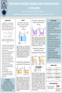
| sciforum-034865 | Cardiolipin regulates select functions of astrocytes dysregulated in chronic neuroinflammatory states | , , | N/A |

|
Show Abstract |
||||||||||||||||||||||||||||||||||||
|
Cardiolipin (CL) is a mitochondrial phospholipid, which has a well-defined role in regulating metabolic processes and maintaining homeostasis. While CL is normally confined to the inner mitochondrial membrane, it may be released extracellularly from damaged or dying cells during physiological and pathological conditions characterized by cellular stress, tissue damage, and cell death. Once released, CL can act as a signalling molecule by interacting with cells in an autocrine and paracrine manner. Previous research demonstrates that extracellular CL regulates select immune functions of microglia, which are the innate immune cells of the brain. However, the effects of CL on astrocyte functions are unknown. Astrocytes support metabolic homeostasis of neurons, facilitate neurotransmission, and are also active participants in neuroimmune responses of the brain. During chronic neuroinflammatory states, astrocytes become over-activated and may cause damage to the surrounding tissues and cells. We hypothesized that extracellular CL modulates the secretion of cytokines and cytotoxins by astrocytes, as well as their phagocytic activity. Therefore, we studied the effects of CL added to the culture media of primary murine astrocytes and human U118 MG astrocytic cells. The phagocytic activity of primary murine astrocytes was measured using fluorescent latex microspheres. The secretion of inflammatory cytokines was monitored by the enzyme-linked immunosorbent assay or immunoblotting. The expression of glial fibrillary acidic protein (GFAP) was measured using immunoblotting. Extracellular CL alone upregulated the phagocytic activity of primary murine astrocytes, as well as the release of monocyte chemoattractant protein (MCP)-1 and interferon (IFN)-β by human U118 MG astrocytic cells. CL inhibited the lipopolysaccharide (LPS)-induced increase in GFAP expression by U118 MG astrocytic cells, as well as the secretion of cytotoxins by the same cell type. Our study demonstrates that extracellular CL regulates select functions of astrocytes, which become dysregulated in chronic neuroinflammatory states, such as those observed in neurodegenerative diseases. |
|||||||||||||||||||||||||||||||||||||||||
| sciforum-034891 | Neurotrophins as intercellular signaling molecules of the brain regulate select immune functions of microglia | , | N/A |

|
Show Abstract |
||||||||||||||||||||||||||||||||||||
|
Microglia are the resident immune cells of the brain, which become activated in response to diverse immune stimuli. Following activation, microglia signal to other central nervous system (CNS) cells by secreting a variety of cytokines. Under specific stimulatory conditions, microglia can also release a spectrum of cytotoxins, including reactive oxygen species (ROS), reactive nitrogen species (RNS), proteases, and other enzymes. Molecules secreted by microglia have specific functions in the neuroimmune response to pathogens and endogenous stimuli. They also contribute to the pathogenesis of neurodegenerative diseases. Neurotrophins (NTs) are proteins that support development, maturation and normal metabolic functions of neurons by interacting with neurotrophin receptors (NTRs) on these cells. Microglia also express several NTRs that signal in response to binding of NTs. These include tropomyosin receptor kinase (Trk)A, TrkB and p75NTR. Binding and activation of these receptors can alter the phenotype, metabolism, and other functions of microglia. We hypothesized that NTR ligands, such as the small molecule ligand LM11A-31 and the NT precursor pro-brain derived neurotrophic factor (proBDNF), bind p75NTR and modulate the immune functions of microglia. We studied the effects of NTR ligands on the following immune functions by using in vitro models of microglia: 1) secretion of cytotoxins and cytokines by lipopolysaccharide (LPS) plus interferon-γ stimulated human THP-1 monocytic cells; 2) release of ROS through the LPS-primed respiratory burst response of dimethyl sulfoxide-differentiated human HL-60 myelomonocytic cells; 3) secretion of nitric oxide by LPS-stimulated murine BV-2 microglia; and 4) phagocytic activity of BV-2 cells. LM11A-31 and proBDNF inhibited the respiratory burst response of differentiated HL-60 microglia-like cells but did not have a significant effect in most other cellular assays. Our data indicate that NTs could serve as intercellular signaling molecules of the CNS by regulating select immune functions of microglia, such as their production of ROS. |
|||||||||||||||||||||||||||||||||||||||||
Event Awards
To acknowledge the support of the conference esteemed authors and recognize their outstanding scientific accomplishments, Cells would like to award the best papers and best speakers as elected by the members of the scientific committee. The Award will consist of 500 Swiss Francs and a free featured paper. We look forward to posting your contributions.


The Awards
Number of Awards Available: 2
The Best Paper Award is given for the paper judged to make the most significant contribution to the conference. There will be two winners selected for this award, the winners will be scholars in an early stage of her career (within seven years of appointment to an independent position)Number of Awards Available: 2
Two best speakers will be selected by the Scientific Committee after evaluation of all selected talks presented during the conference. The winners will be selected from the videos uploaded through our submission system.Sponsors and Partners
For information regarding sponsorship and exhibition opportunities, please click here.
Organizers
B. Exosomes and Extracellular Vesicles in Health and Disease
Show all published submissions (2) Hide published submissions (2)
Submissions
List of Papers (2) Toggle list
D. The cross talk between cell death regulation and metabolism
Show all published submissions (1) Hide published submissions (1)
Submissions
List of Papers (1) Toggle list
F. Signaling and Metabolic Cross-talk in the Tumor Microenvironment
Session Chair
Professor Danny N. Dhanasekaran, Stephenson Cancer Center, The University of Oklahoma Health Sciences Center, OKC (US)
Show all published submissions (1) Hide published submissions (1)
Submissions
List of Papers (1) Toggle list
G. The cross-talk between immune cells and tissue microenvironment
Show all accepted abstracts (1) Hide accepted abstracts (1)
List of Accepted Abstracts (1) Toggle list
P. Posters
Show all published submissions (3) Hide published submissions (3)
Submissions
List of Papers (3) Toggle list


