Event submissions
Published
This submission belongs to the session S7. Pulmonology of the event The 3rd International Online Conference on Clinical Medicine
Published date
12 Nov, 2025
Academic Editor
'%3e%3cpath%20d='M12.6657%2010.9104C12.4185%2010.325%2012.0599%209.79327%2011.6097%209.34482C11.1609%208.89508%2010.6293%208.5365%2010.0442%208.28888C10.0389%208.28626%2010.0337%208.28495%2010.0285%208.28233C10.8446%207.69279%2011.3752%206.73249%2011.3752%205.64904C11.3752%203.8542%209.92103%202.39999%208.1262%202.39999C6.33136%202.39999%204.87715%203.8542%204.87715%205.64904C4.87715%206.73249%205.40774%207.69279%206.22393%208.28364C6.21869%208.28626%206.21345%208.28757%206.20821%208.29019C5.62129%208.5378%205.09463%208.89284%204.64265%209.34613C4.1929%209.79493%203.83432%2010.3266%203.58671%2010.9117C3.34345%2011.4845%203.21226%2012.0987%203.20023%2012.7209C3.19988%2012.7349%203.20233%2012.7488%203.20744%2012.7619C3.21255%2012.7749%203.22022%2012.7867%203.22998%2012.7968C3.23975%2012.8068%203.25142%2012.8147%203.26431%2012.8202C3.2772%2012.8256%203.29105%2012.8284%203.30504%2012.8284H4.09109C4.14874%2012.8284%204.19459%2012.7825%204.1959%2012.7262C4.2221%2011.7148%204.62823%2010.7676%205.34617%2010.0497C6.08899%209.30683%207.0755%208.89808%208.1262%208.89808C9.17689%208.89808%2010.1634%209.30683%2010.9062%2010.0497C11.6242%2010.7676%2012.0303%2011.7148%2012.0565%2012.7262C12.0578%2012.7838%2012.1037%2012.8284%2012.1613%2012.8284H12.9474C12.9613%2012.8284%2012.9752%2012.8256%2012.9881%2012.8202C13.001%2012.8147%2013.0126%2012.8068%2013.0224%2012.7968C13.0322%2012.7867%2013.0398%2012.7749%2013.0449%2012.7619C13.0501%2012.7488%2013.0525%2012.7349%2013.0522%2012.7209C13.0391%2012.0947%2012.9094%2011.4855%2012.6657%2010.9104ZM8.1262%207.9024C7.52486%207.9024%206.9589%207.6679%206.53312%207.24211C6.10733%206.81633%205.87283%206.25037%205.87283%205.64904C5.87283%205.0477%206.10733%204.48174%206.53312%204.05596C6.9589%203.63018%207.52486%203.39567%208.1262%203.39567C8.72753%203.39567%209.29349%203.63018%209.71927%204.05596C10.1451%204.48174%2010.3796%205.0477%2010.3796%205.64904C10.3796%206.25037%2010.1451%206.81633%209.71927%207.24211C9.29349%207.6679%208.72753%207.9024%208.1262%207.9024Z'%20fill='%235D1EE1'/%3e%3c/g%3e%3c/svg%3e) Sukhwinder Sohal
Sukhwinder SohalCitation
Eugene Y.H. Yeung, Pulmonary Aspergillosis in a Patient with Diabetic Ketoacidosis, in Proceedings of The 3rd International Online Conference on Clinical Medicine, 17 November–19 November 2025, MDPI: Basel, Switzerland
Share
Email
Facebook
Twitter
LinkedIn
Pulmonary Aspergillosis in a Patient with Diabetic Ketoacidosis
'%3e%3cpath%20d='M12.6647%2010.9104C12.4176%2010.325%2012.0589%209.7933%2011.6088%209.34485C11.16%208.89511%2010.6283%208.53653%2010.0432%208.28891C10.038%208.28629%2010.0327%208.28498%2010.0275%208.28236C10.8437%207.69282%2011.3743%206.73252%2011.3743%205.64907C11.3743%203.85423%209.92005%202.40002%208.12522%202.40002C6.33038%202.40002%204.87618%203.85423%204.87618%205.64907C4.87618%206.73252%205.40677%207.69282%206.22296%208.28367C6.21772%208.28629%206.21248%208.2876%206.20724%208.29022C5.62031%208.53783%205.09365%208.89287%204.64167%209.34616C4.19192%209.79496%203.83334%2010.3266%203.58573%2010.9117C3.34247%2011.4846%203.21128%2012.0987%203.19925%2012.721C3.1989%2012.735%203.20135%2012.7489%203.20646%2012.7619C3.21157%2012.7749%203.21924%2012.7868%203.22901%2012.7968C3.23877%2012.8068%203.25045%2012.8148%203.26334%2012.8202C3.27623%2012.8256%203.29007%2012.8284%203.30406%2012.8284H4.09012C4.14776%2012.8284%204.19362%2012.7825%204.19493%2012.7262C4.22113%2011.7148%204.62726%2010.7676%205.34519%2010.0497C6.08802%209.30686%207.07452%208.89811%208.12522%208.89811C9.17592%208.89811%2010.1624%209.30686%2010.9052%2010.0497C11.6232%2010.7676%2012.0293%2011.7148%2012.0555%2012.7262C12.0568%2012.7839%2012.1027%2012.8284%2012.1603%2012.8284H12.9464C12.9604%2012.8284%2012.9742%2012.8256%2012.9871%2012.8202C13%2012.8148%2013.0117%2012.8068%2013.0214%2012.7968C13.0312%2012.7868%2013.0389%2012.7749%2013.044%2012.7619C13.0491%2012.7489%2013.0515%2012.735%2013.0512%2012.721C13.0381%2012.0947%2012.9084%2011.4856%2012.6647%2010.9104ZM8.12522%207.90243C7.52388%207.90243%206.95792%207.66793%206.53214%207.24215C6.10636%206.81636%205.87185%206.2504%205.87185%205.64907C5.87185%205.04773%206.10636%204.48177%206.53214%204.05599C6.95792%203.63021%207.52388%203.3957%208.12522%203.3957C8.72655%203.3957%209.29252%203.63021%209.7183%204.05599C10.1441%204.48177%2010.3786%205.04773%2010.3786%205.64907C10.3786%206.2504%2010.1441%206.81636%209.7183%207.24215C9.29252%207.66793%208.72655%207.90243%208.12522%207.90243Z'%20fill='%235D1EE1'/%3e%3c/g%3e%3c/svg%3e)
Eugene Y.H. Yeung 1,2,3
1. Department of Pathology and Laboratory Medicine, Faculty of Medicine, University of British Columbia, Vancouver, British Columbia V5Z 1M9, Canada, Canada
2. Continuing Pharmacy Professional Development, Faculty of Pharmaceutical Sciences, University of British Columbia, Vancouver, British Columbia V6T 1Z3, Canada
3. Clinical Faculty, School of Medicine, Simon Fraser University, Surrey, British Columbia V3T 0A3, Canada
Abstract
Introduction
Pulmonary mucormycosis is a potential complication in patients with diabetic ketoacidosis. Isolation of filamentous fungus from bronchoscopy specimens of this population may prompt clinicians to include pulmonary mucormycosis in their differential diagnoses and initiate high-dose intravenous liposomal amphotericin (>5 mg/kg/day). Amphotericin is an expensive therapy and is frequently associated with adverse effects such as nephrotoxicity, phlebitis, and acute febrile reaction. We encountered a case in which pulmonary aspergillosis rather than mucormycosis was diagnosed in a patient with diabetic ketoacidosis.
Method
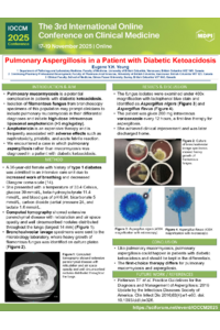
A 36-year-old female with history of type 1 diabetes was admitted to an intensive care unit due to increased work of breathing and decreased Glasgow coma scale (14). She presented with a temperature of 33.4 Celsius, glucose 39 mmol/L, beta-hydroxybutyrate 11.4 mmol/L, and blood gas of pH 6.94, bicarbonate 5 mmol/L, carbon dioxide partial pressure 24, and lactate 1.6 mmol/L. Computed tomography showed extensive parenchymal disease with reticulation and air space opacity and well circumscribed nodules distributed throughout the lungs (largest 14 mm). (Figure 1 https://drive.google.com/file/d/16_Tw4Uoa2FbavqeM5_u7IomSOTeoMOo0/view?usp=sharing). Bronchoalveolar lavage specimens were sent to the microbiology laboratory, where heavy growth of filamentous fungus was identified on culture plates (Figure 2 https://drive.google.com/file/d/1CSIng6R2YYDfAt9evfVnVm6zvLspKvvV/view?usp=sharing).
Results
The fungus isolates were examined under 400x magnification with lactophenol blue stain and identified as Aspergillus nigers (Figure 3 https://drive.google.com/file/d/1Qjd-CCyIusivBVLfX0uTc2GsijpMp0oV/view?usp=sharing) and Aspergillus flavus (Figure 4 https://drive.google.com/file/d/1-4nY7_RInykv-HWUXuGF-IZ-8VT-E_ra/view?usp=sharing). The patient was given 200 mg intravenous voriconazole every 12 hours, a first-line therapy for aspergillosis. She achieved clinical improvement and was later discharged home.
Conclusion
Like pulmonary mucormycosis, pulmonary aspergillosis could happen in patients with diabetic ketoacidosis and should be kept in the differentials. The first-choice therapy differs for pulmonary mucormycosis and aspergillosis.
Keywords
Pulmonary mucormycosis
Aspergillosis
Aspergillus nigers
diabetic ketoacidosis
Aspergillus flavus
Poster
135989 .pdf
Factors associated with higher respiratory disturbance index than apnea–hyponea index in patients with obstructive sleep apnea
Individual Patient Data Meta-Analysis of Histopathological Patterns and Vaping-Associated Lung Injury: Different Pathophysiology of Nicotine and Marijuana-Based Vaping