Event submissions
Published

This submission belongs to the session F. Nanomedicine and Nanobiotechnology of the event The 2nd International Online Conference on Nanomaterials
Published date
11 Nov, 2020
Citation
Miruna S. Stan, Ionela Cristina Nica, Julliette Moreau, Maité Callewaert, Françoise Chuburu, Anca Dinischiotu, Sorina Nicoleta Voicu, Hildegard Herman, Anca Hermenean, Cyril Cadiou, Fluorescent chitosan nanogels developed for targeting endothelial cells of axillary lymph nodes, in Proceedings of The 2nd International Online Conference on Nanomaterials, 15 November–30 November 2020, MDPI: Basel, Switzerland, doi: 10.3390/IOCN2020-07847
Share
Email
Facebook
Twitter
LinkedIn
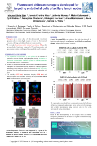
Fluorescent chitosan nanogels developed for targeting endothelial cells of axillary lymph nodes

Miruna S. Stan 1

Ionela Cristina Nica 2
'%3e%3cpath%20d='M12.6647%2010.9104C12.4176%2010.325%2012.0589%209.7933%2011.6088%209.34485C11.16%208.89511%2010.6283%208.53653%2010.0432%208.28891C10.038%208.28629%2010.0327%208.28498%2010.0275%208.28236C10.8437%207.69282%2011.3743%206.73252%2011.3743%205.64907C11.3743%203.85423%209.92005%202.40002%208.12522%202.40002C6.33038%202.40002%204.87618%203.85423%204.87618%205.64907C4.87618%206.73252%205.40677%207.69282%206.22296%208.28367C6.21772%208.28629%206.21248%208.2876%206.20724%208.29022C5.62031%208.53783%205.09365%208.89287%204.64167%209.34616C4.19192%209.79496%203.83334%2010.3266%203.58573%2010.9117C3.34247%2011.4846%203.21128%2012.0987%203.19925%2012.721C3.1989%2012.735%203.20135%2012.7489%203.20646%2012.7619C3.21157%2012.7749%203.21924%2012.7868%203.22901%2012.7968C3.23877%2012.8068%203.25045%2012.8148%203.26334%2012.8202C3.27623%2012.8256%203.29007%2012.8284%203.30406%2012.8284H4.09012C4.14776%2012.8284%204.19362%2012.7825%204.19493%2012.7262C4.22113%2011.7148%204.62726%2010.7676%205.34519%2010.0497C6.08802%209.30686%207.07452%208.89811%208.12522%208.89811C9.17592%208.89811%2010.1624%209.30686%2010.9052%2010.0497C11.6232%2010.7676%2012.0293%2011.7148%2012.0555%2012.7262C12.0568%2012.7839%2012.1027%2012.8284%2012.1603%2012.8284H12.9464C12.9604%2012.8284%2012.9742%2012.8256%2012.9871%2012.8202C13%2012.8148%2013.0117%2012.8068%2013.0214%2012.7968C13.0312%2012.7868%2013.0389%2012.7749%2013.044%2012.7619C13.0491%2012.7489%2013.0515%2012.735%2013.0512%2012.721C13.0381%2012.0947%2012.9084%2011.4856%2012.6647%2010.9104ZM8.12522%207.90243C7.52388%207.90243%206.95792%207.66793%206.53214%207.24215C6.10636%206.81636%205.87185%206.2504%205.87185%205.64907C5.87185%205.04773%206.10636%204.48177%206.53214%204.05599C6.95792%203.63021%207.52388%203.3957%208.12522%203.3957C8.72655%203.3957%209.29252%203.63021%209.7183%204.05599C10.1441%204.48177%2010.3786%205.04773%2010.3786%205.64907C10.3786%206.2504%2010.1441%206.81636%209.7183%207.24215C9.29252%207.66793%208.72655%207.90243%208.12522%207.90243Z'%20fill='%235D1EE1'/%3e%3c/g%3e%3c/svg%3e)
Julliette Moreau 3
Maité Callewaert 3
Cyril Cadiou 3
Françoise Chuburu 3
Hildegard Herman 4

Anca Hermenean 4

Anca Dinischiotu 2
Sorina Nicoleta Voicu 2
1. Departament of Biochemistry and Molecular Biology, Faculty of Biology, University of Bucharest, 91-95 Splaiul Independentei, 050095 Bucharest, Romania
2. Department of Biochemistry and Molecular Biology, Faculty of Biology, University of Bucharest, 91-95 Splaiul Independentei, 050095 Bucharest, Romania
3. Institut de Chimie Moléculaire de Reims, CNRS UMR 7312, University of Reims Champagne-Ardenne URCA, 51685 Reims Cedex 2, France
4. Aurel Ardelean Institute of Life Sciences, Vasile Godis Western University of Arad, Rebreanu 86, 310414 Arad, Romania
Abstract
Nanogels are a novel class of three-dimensional cross-linked polymers able to retain high amounts of water in their network structure, with large potential applications in nanomedicine. In our study, the polymer matrix selected was chitosan, as this polysaccharide biopolymer composed of N-acetylglucosamine and glucosamine residues exhibit great biocompatibility and low toxicity. The preparation was performed by ionic gelation in the presence of hyaluronic acid and sodium tripolyphosphate, having rhodamine or fluorescein isothiocyanate molecules grafted on chitosan backbone. In order to validate the possible usage of these chitosan-fluorophores conjugates for fluorescence imaging purposes in cancer diagnostics and therapy, their biological effect was assessed on SVEC4-10 cells (a simian virus 40-transformed mouse microvascular endothelial cell line). Cell viability, membrane integrity and nanogels uptake were examined following the exposure for 6 and 24 hours at concentrations up to 120 µg/mL. A good biocompatibility was obtained after both time intervals of incubation with nanogels, as no increase in cell death or membrane damage being noticed compared to control. By examination on confocal laser scanning microscopy, the both types of fluorescent nanogels agglomerated on the surface of cell membrane, their cellular internalization being observed only for few cells, preferentially at the cell periphery. In conclusion, based on the biocompatibility of the nanogels, these can further incorporate gadolinium for an improved magnetic resonance Imaging effect in nanomedicine.
Acknowledgments This work was supported by a grant of the Romanian Ministry of Research and Innovation, CCCDI - UEFISCDI, project number PN-III-P3-3.1-PM-RO-FR-2019-0204 / 6 BM/ 2019, within PNCDI III.
Keywords
chitosan
nanoparticles
lymph nodes
medical imaging
Manuscript
sciforum-039217-done.pdf
Poster
Poster - MS Stan.pdf
Removal of Manganese using Polymer gel composites
Formation, Phase Composition and Memristive Properties of Titanium Oxide Nanodots