Event submissions
Published

This submission belongs to the session S3. The Biology of Natural Products in Disease Pathophysiology: Mechanisms of Action of the event The 1st International E-Conference on Antioxidants in Health and Disease
Published date
01 Dec, 2020
Citation
Danila Cianciosi, The preliminary effect of Manuka honey on cancer stem-like cells from colonspheres, in Proceedings of The 1st International E-Conference on Antioxidants in Health and Disease, 1 December–15 December 2020, MDPI: Basel, Switzerland, doi: 10.3390/CAHD2020-08636
Share
Email
Facebook
Twitter
LinkedIn
The preliminary effect of Manuka honey on cancer stem-like cells from colonspheres

Danila Cianciosi 1
1. Università Politecnica delle Marche, Italy
Abstract
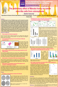
Honey has always been used not only as a food/sweetener but also as a medicine since ancient times. The quality and composition depend on many factors, including the botanical origin, the environmental, processing and storage conditions[1]. The nutritional characteristics and the preventive/therapeutic effect of honey are due to its composition: it contains over 180 different types of compounds including water, carbohydrates, enzymes, amino acids, minerals, vitamins and different phytochemicals[2] . Among the different effects on health, those most studied in the scientific literature are its antibacterial activity[3] and the antioxidant capacity[4]. In recent years the potential anticancer effect of honey, in several tumor cell lines, has also been a reason of study[5]. Among different types of honey, Manuka, has shown a high anticancer effect, especially in colon cancer cells (LoVo and HCT-116)[6,7] while it is little known on its effect in cancer stem cells' (CSCs; a rare population of cells within the tumor mass that seem to be responsible for the tumor onset) chemoresistance and the presence of relapse [8]. Therefore, the effect of Manuka honey on CSCs-like was evaluated. In general, CSCs-like were enriched from the monolayer population of HCT-116 through the in vitro sphere forming assay[9]. This honey was able to modify the morphological parameters of the spheroids, reducing the size and volume of the entire culture. The treatment of CSCs-like enriched colonospheres with Manuka honey also led to an intracellular accumulation of ROS and induction of apoptosis. Furthermore, through real-time PCR, down-regulation of ABCG2 gene expression (one of the efflux pumps closely associated with the chemoresistance phenotype) was observed.
References
- DOI: 10.1016/j.lwt.2017.08.079
- DOI: 10.3390/molecules23092322
- DOI: 10.1155/2019/2464507
- DOI: 10.1016/j.jff.2016.05.008
- DOI: 10.1016/j.clnu.2018.12.019
- DOI: 10.1039/c8fo00164b
- DOI: 10.1039/c8fo00165k
- DOI: 10.1016/j.phrs.2018.08.006
- DOI: 10.18632/oncotarget.6261
Keywords
honey
cancer
cancer stem cells
polyphenols
spheroids
3d
Manuscript
The preliminary effect of Manuka_Cianciosi.pdf
Poster
sciforum-037467.pdf
A new role of red wine in modulating erythrocytes antioxidant defense
Evaluation of the phenolic profile, mineral, and fatty acid content and antioxidant activity of Black cumin before and after an in vitro simulated gastrointestinal digestion.