Event submissions
Published

This submission belongs to the session E. Technological Advances in Metabolomics (including Bioinformatics) of the event The 2nd International Electronic Conference on Metabolomics
Published date
20 Nov, 2017
Citation
Christian Metallo, Tracing Compartment-Specific Redox Pathways Using Stable Isotopes and Mass Spectrometry, in Proceedings of The 2nd International Electronic Conference on Metabolomics, 20 November–27 November 2017, MDPI: Basel, Switzerland, doi: 10.3390/iecm-2-05000
Share
Email
Facebook
Twitter
LinkedIn
Tracing Compartment-Specific Redox Pathways Using Stable Isotopes and Mass Spectrometry
'%3e%3cpath%20d='M12.6647%2010.9104C12.4176%2010.325%2012.0589%209.7933%2011.6088%209.34485C11.16%208.89511%2010.6283%208.53653%2010.0432%208.28891C10.038%208.28629%2010.0327%208.28498%2010.0275%208.28236C10.8437%207.69282%2011.3743%206.73252%2011.3743%205.64907C11.3743%203.85423%209.92005%202.40002%208.12522%202.40002C6.33038%202.40002%204.87618%203.85423%204.87618%205.64907C4.87618%206.73252%205.40677%207.69282%206.22296%208.28367C6.21772%208.28629%206.21248%208.2876%206.20724%208.29022C5.62031%208.53783%205.09365%208.89287%204.64167%209.34616C4.19192%209.79496%203.83334%2010.3266%203.58573%2010.9117C3.34247%2011.4846%203.21128%2012.0987%203.19925%2012.721C3.1989%2012.735%203.20135%2012.7489%203.20646%2012.7619C3.21157%2012.7749%203.21924%2012.7868%203.22901%2012.7968C3.23877%2012.8068%203.25045%2012.8148%203.26334%2012.8202C3.27623%2012.8256%203.29007%2012.8284%203.30406%2012.8284H4.09012C4.14776%2012.8284%204.19362%2012.7825%204.19493%2012.7262C4.22113%2011.7148%204.62726%2010.7676%205.34519%2010.0497C6.08802%209.30686%207.07452%208.89811%208.12522%208.89811C9.17592%208.89811%2010.1624%209.30686%2010.9052%2010.0497C11.6232%2010.7676%2012.0293%2011.7148%2012.0555%2012.7262C12.0568%2012.7839%2012.1027%2012.8284%2012.1603%2012.8284H12.9464C12.9604%2012.8284%2012.9742%2012.8256%2012.9871%2012.8202C13%2012.8148%2013.0117%2012.8068%2013.0214%2012.7968C13.0312%2012.7868%2013.0389%2012.7749%2013.044%2012.7619C13.0491%2012.7489%2013.0515%2012.735%2013.0512%2012.721C13.0381%2012.0947%2012.9084%2011.4856%2012.6647%2010.9104ZM8.12522%207.90243C7.52388%207.90243%206.95792%207.66793%206.53214%207.24215C6.10636%206.81636%205.87185%206.2504%205.87185%205.64907C5.87185%205.04773%206.10636%204.48177%206.53214%204.05599C6.95792%203.63021%207.52388%203.3957%208.12522%203.3957C8.72655%203.3957%209.29252%203.63021%209.7183%204.05599C10.1441%204.48177%2010.3786%205.04773%2010.3786%205.64907C10.3786%206.2504%2010.1441%206.81636%209.7183%207.24215C9.29252%207.66793%208.72655%207.90243%208.12522%207.90243Z'%20fill='%235D1EE1'/%3e%3c/g%3e%3c/svg%3e)
Christian Metallo 1
1. University of California, San Diego, Department of Bioengineering
Abstract
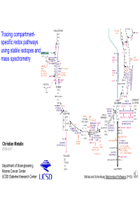
Metabolism is central to virtually all cellular functions and contributes to a range of diseases. A quantitative understanding of how biochemical pathways are dysregulated in the context of diseases such as cancer and metabolic syndrome is necessary to identify new therapeutic targets. To this end we apply stable isotope tracers, mass spectrometry, and metabolic flux analysis (MFA) to study metabolism in mammalian cells, animal models, and human patients. Using these approaches we have characterized how proliferating and differentiated cells regulate flux of glucose and amino acids into mitochondria for maintaining redox homeostasis and lipid biosynthesis. Recently, we have developed novel methods for studying pyridine nucleotide metabolism, employing 2H tracers and mass spectrometry to quantify how specific metabolic pathways are used to regenerate NADH and NADPH. To better understand how redox pathways are regulated in the cytosol and mitochondrial matrix we have generated compartment-specific enzyme reporters that exploit the neomorphic activity of mutant isocitrate dehydrogenases (IDHs). Specifically, R132H IDH1 and R172K IDH2 produce (D)2-hydroxyglutarate (2HG) in the cytosol and mitochondria, respectively. Quantitation of labeling from specifically labeled 2H tracers provides critical insights into NAD(P)H-producing pathways in each compartment. We have employed this approach to identify redox pathway regulation under hypoxia, where oxidative pentose phosphate pathway flux is upregulated to fuel reductive carboxylation. The application of MFA to cell and animal models greatly improves our ability to characterize intracellular metabolic processes, providing a mechanistic understanding of cellular physiology and metabolic function.
Keywords
metabolic flux analysis
NADPH
NADH
mitochondria
cytosol
IDH1
hypoxia
serine
one carbon metabolism
Poster
Metallo_IECM_Nov_2017.pdf
Investigation of Salinity Tolerance Mechanism in Barley Roots Using Semi-Targeted Lipidomics Approach with High-Resolution Mass Spectrometry Techniques
Metabolomics in Chronobiology: Metabolic Alterations by Sleep Loss and Circadian Function