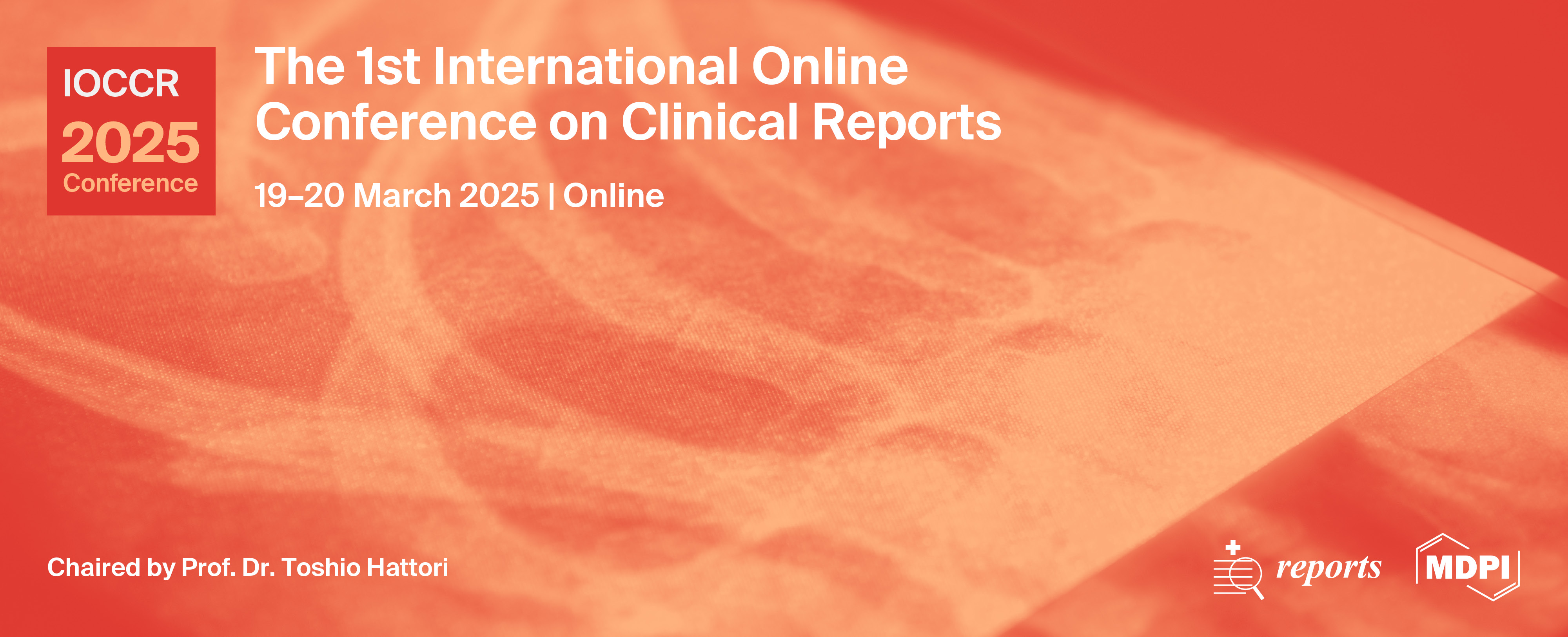
The 1st International Online Conference on Clinical Reports
Thank you for attending IOCCR 2025!
On behalf of the conference organizing committee of The 1st International Online Conference on Clinical Reports (IOCCR 2025), we would like to express our appreciation to all of the participants for their contributions. From all accounts, the event was a success.
The IOCCR 2025 Best Oral Presentation Awards and Best Poster Awards have been announced in the Award Winners Announcement section.
Click HERE for the Certificate of Participation.
Click HERE for the Book of Abstracts.
Click HERE for the Poster Gallery.
Publication Opportunities for IOCCR 2025 Participants.
For any inquiries, please contact us at ioccr@mdpi.com.
the chair
We are delighted to welcome you to the 1st International Online Conference on Clinical Reports (IOCCR 2025), which will be held online and promoted by the open access MDPI journal Reports (ISSN: 2571-841X; IF: 0.8).
This conference offers a platform for doctors, experts, and medical-related practitioners in the field of medical cases, images, and videos in human medicine to engage in an exchange of ideas and share cutting-edge research findings. The conference will encompass diverse facets of clinical medicine, including, but not limited to, the following:
1. Disaster/Climate Change Medicine;
2. Cancer;
3. Cardiovascular Diseases;
4. Oral Diseases;
5. Orthopedic Surgery.
We are excited to have this opportunity to explore the latest developments and future trends in clinical medicine alongside related practitioners from around the world. This conference will serve as a valuable platform for exchange and cooperation, fostering the development and progression of clinical medicine.
The conference will be held entirely online, allowing people from all around the world to participate without any concerns about travel or related expenses. An online conference offers a platform for quick and direct exchanges of the latest research findings and innovative ideas.
Lastly, we would like to express our sincere gratitude for your unwavering support and participation and wish this conference great success!
Kind regards,
Prof. Dr. med. Toshio Hattori
Director, Rouken Nursing Home Akane, Japan
Graduate School of Public Health, Shizuoka Graduate University of Public Health, Japan
Emeritus Professor, Tohoku University
Meet the Event Chair
Important Dates

- Abstract submission deadlineJan 10, 2025
- Abstract acceptance notificationFeb 10, 2025
- Registration end dateMar 17, 2025
Meet Our Speakers
Dr. Ryo Naito
Department of Cardiovascular Biology and Medicine, Juntendo University Graduate School of Medicine, Tokyo, Japan, Cardiovascular Respiratory Sleep Medicine, Juntendo University Graduate School of Medicine, Tokyo, Japan;
Ryo Naito is an Associate Professor of Department of Cardiovascular Biology and Medicine, Juntendo University, Tokyo, Japan. His research program has three main components: prevention of cardiovascular disease, cardiac rehabilitation, and sleep apnea.
He received his MD degree from Juntendo University in 2005 and completed a 2-year residency in Internal Medicine at Toranomon Hospital, Tokyo, Japan. He earned a Master of Science (MSc) from McMaster University, Ontario, Canada and a PhD degree from Juntendo University.
Dr. Monica Casiraghi
Division of Thoracic Surgery, IEO, European Institute of Oncology IRCCS, and Department of Oncology and Haemato-Oncology, University of Milan, Italy;
Dr. Casiraghi is Deputy Director of the Division of Thoracic Surgery at European Institute of Oncology in Milan and Assistant Professor of the Department of Oncology and Hemato-oncology at the University of Milan, Italy. Since 2008, she worked at European Institute of Oncology in Milan where she became expert in thoracic oncology and minimally invasive techniques such as robotic surgery. She is the PI and co-PI in different projects and working group both for lung cancer, mesothelioma and thymoma treatments, and she is recently focusing her research on microbiome characterization both for the early detection of lung cancer and for more advance stage NSCLC treated with immunotherapy. She is the first author and co-author of many papers in peer reviewed international journals. Dr. Casiraghi is editor and reviewer of international journals. She is actually board member of the Thoracic Domain and chair of the Surgical Oncology task force of EACTS, and member of ESTS Robotic working group and Women in General Thoracic Surgery.
Prof. Dr. Shoji Yokobori
Department of Emergency and Critical Care Medicine, Nippon Medical School Graduate School of Medicine, Japan;
Shoji Yokobori, MD PhD is the Professor, Director and Chair of Department of Emergency and Critical Care Medicine, Nippon Medical School and Graduate School of Nippon Medical School. He graduated from Gunma University of Medicine in 1999 and obtained a Ph.D from Graduate School of Nippon Medical School in 2005. He had carried out research on neurotrauma as a visiting researcher at University of Miami Miller School of Medicine, USA from 2010 to 2013. His specialty is neurotauma and neurosurgical emergency. He is currently engaged in clinical works, and also in research on basic experiments of nerve regeneration, transplantation of stem cells, brain hypothermia and heatstroke.
Prof. Dr. Manabu Fukumoto
International Research Institute of Disaster Science (IRIDeS), Tohoku University, Miyagi, Japan;
Professor Manabu Fukumoto received his Phd in 1981 from Kyoto University, Japan and got his Pathologist certification in 1982 from Japanese Pathological Association. He is a Specially Appointed Professor & Professor Emeritus at Tokyo Medical University and Tohoku University and a Visiting Professor at the International Research Institute of Disaster Science, Tohoku University. He is also working as a Senior Visiting Scientist at RIKEN Center for Advanced Intelligence Project, Japan. His Research Activities are focused on the Efffects of the Fukushima Daiichi Nuclear Power Plant accident on the ecosystem.
Prof. Dr. Hiroshi Inui
Saitama Medical Center, Saitama Medical University, Japan;
Graduated from University of Tokyo in 2001,
2001~ Tokyo University Hospital (resident doctor),
2016~Tokyo University Hospital (director of the joint replacement center),
2023.6~ Saitama Medical University, Saitama Medical center (Professor)
Prof. Dr. Roberto Lo Giudice
Department of Human Pathology of adults and developmental age, Messina University, Messina, Italy;
Graduated cum laude at Messina University (Italy), and moved to Federico II University, Naples (Italy) to specialize in Oral Surgery, where he graduated cum laude. PhD in the Department of Biomedical, Clinical, and Experimental Sciences (XXXII course) at Messina University. Post-doc researcher in the Department of Human Pathology in Adulthood and Childhood at Messina University. Private dentist at Studio Odontoiatrico Lo Giudice Messina, Italy, since 2012. Attended an advanced course in oral surgery with piezoelectric devices and an advanced course in Implant surgery and surgical anatomy. Winner of the Young Podium Competition in 2014 in Tirana (Albany), 2015 Mahadia (Tunisia), and 2015 Estoril (Portugal). Winner of best poster at 2016 Collegio Docenti Rome (Italy). Visiting researcher at the Vita Salute S. Raffaele University (Milan), Department of Oral Surgery and Implantology, in 2017. Visiting researcher at the University Hospital of Geneva, Department of Maxillo-facial Surgery and Oral Surgery, in 2018. Founder and component of the board of the intHEMA (International Non-transfusional Hemocomponents Academy). National Secretary of the SIdCO (Italian Society of Oral Surgery) (2017-2019).
- S1. Disaster/Climate Change Medicine
- S2. Cancer
- S3. Cardiovascular Diseases
- S4. Oral Diseases
- S5. Orthopedic Surgery
Sponsors and Partners
Organizer


Media partner


On behalf of the chairs of IOCCR 2025, we are pleased to announce the winners of the Best Oral Presentation Awards and Best Poster Awards. Congratulations! We will be contacting the awarded Oral / Poster presenters on the next steps.
The Best Oral Presentation Awards have been awarded to:
|
ID |
Title |
Authors |
|
Improving bone protection provision for patients with fragility fractures |
Aaron Goldberg |
|
| sciforum-111519 | Proteolyzed but not full-length galectin-9 in plasma predicts the prognosis of COVID-19 | Hiroko Iwasaki-Hozumi, Toshiro Niki, Takashi Matsuba, Yosuke Maeda, Toshio Hattori |
The Best Poster Awards have been awarded to:
|
ID |
Title |
Authors |
| sciforum-110879 | Paternal Teratogen Exposure and Risk of Congenital Heart Disease in Offspring: A Two-Year Retrospective Observational Study | Shadab Ahamad, Paramvir Singh, Anuj Kumar Sharma, Adhi Arya, Prachi Kukshal |
DAY 1
Session 3. Cardiovascular Diseases
Session 2. Cancer
DAY 2
Session 1. Disaster/Climate Change Medicine
Session 5. Orthopedic Surgery
Session 4. Oral Diseases
Ms. Sunny Shu
Ms. Adelina Platon
Email: ioccr@mdpi.com
For inquiries regarding submissions and sponsorship opportunities, please feel free to contact us.