Event submissions
Published
This submission belongs to the session S2. Hydrogels, Organogels, Xerogels, and Aerogels of the event The 1st International Online Conference on Gels
Published date
28 Nov, 2025
Academic Editor
'%3e%3cpath%20d='M12.6657%2010.9104C12.4185%2010.325%2012.0599%209.79327%2011.6097%209.34482C11.1609%208.89508%2010.6293%208.5365%2010.0442%208.28888C10.0389%208.28626%2010.0337%208.28495%2010.0285%208.28233C10.8446%207.69279%2011.3752%206.73249%2011.3752%205.64904C11.3752%203.8542%209.92103%202.39999%208.1262%202.39999C6.33136%202.39999%204.87715%203.8542%204.87715%205.64904C4.87715%206.73249%205.40774%207.69279%206.22393%208.28364C6.21869%208.28626%206.21345%208.28757%206.20821%208.29019C5.62129%208.5378%205.09463%208.89284%204.64265%209.34613C4.1929%209.79493%203.83432%2010.3266%203.58671%2010.9117C3.34345%2011.4845%203.21226%2012.0987%203.20023%2012.7209C3.19988%2012.7349%203.20233%2012.7488%203.20744%2012.7619C3.21255%2012.7749%203.22022%2012.7867%203.22998%2012.7968C3.23975%2012.8068%203.25142%2012.8147%203.26431%2012.8202C3.2772%2012.8256%203.29105%2012.8284%203.30504%2012.8284H4.09109C4.14874%2012.8284%204.19459%2012.7825%204.1959%2012.7262C4.2221%2011.7148%204.62823%2010.7676%205.34617%2010.0497C6.08899%209.30683%207.0755%208.89808%208.1262%208.89808C9.17689%208.89808%2010.1634%209.30683%2010.9062%2010.0497C11.6242%2010.7676%2012.0303%2011.7148%2012.0565%2012.7262C12.0578%2012.7838%2012.1037%2012.8284%2012.1613%2012.8284H12.9474C12.9613%2012.8284%2012.9752%2012.8256%2012.9881%2012.8202C13.001%2012.8147%2013.0126%2012.8068%2013.0224%2012.7968C13.0322%2012.7867%2013.0398%2012.7749%2013.0449%2012.7619C13.0501%2012.7488%2013.0525%2012.7349%2013.0522%2012.7209C13.0391%2012.0947%2012.9094%2011.4855%2012.6657%2010.9104ZM8.1262%207.9024C7.52486%207.9024%206.9589%207.6679%206.53312%207.24211C6.10733%206.81633%205.87283%206.25037%205.87283%205.64904C5.87283%205.0477%206.10733%204.48174%206.53312%204.05596C6.9589%203.63018%207.52486%203.39567%208.1262%203.39567C8.72753%203.39567%209.29349%203.63018%209.71927%204.05596C10.1451%204.48174%2010.3796%205.0477%2010.3796%205.64904C10.3796%206.25037%2010.1451%206.81633%209.71927%207.24211C9.29349%207.6679%208.72753%207.9024%208.1262%207.9024Z'%20fill='%235D1EE1'/%3e%3c/g%3e%3c/svg%3e) SIDI A. BENCHERIF
SIDI A. BENCHERIFCitation
Ali Mahnavi, Arjan Atwal, Ava Carroll, Ying Yang, Pooya Davoodi, Nanofibrous Hyaluronic Acid/Gelatin Methacrylate Hydrogels Enhance Cartilage Regeneration, in Proceedings of The 1st International Online Conference on Gels, 3 December–5 December 2025, MDPI: Basel, Switzerland
Share
Email
Facebook
Twitter
LinkedIn
Nanofibrous Hyaluronic Acid/Gelatin Methacrylate Hydrogels Enhance Cartilage Regeneration
'%3e%3cpath%20d='M12.6647%2010.9104C12.4176%2010.325%2012.0589%209.7933%2011.6088%209.34485C11.16%208.89511%2010.6283%208.53653%2010.0432%208.28891C10.038%208.28629%2010.0327%208.28498%2010.0275%208.28236C10.8437%207.69282%2011.3743%206.73252%2011.3743%205.64907C11.3743%203.85423%209.92005%202.40002%208.12522%202.40002C6.33038%202.40002%204.87618%203.85423%204.87618%205.64907C4.87618%206.73252%205.40677%207.69282%206.22296%208.28367C6.21772%208.28629%206.21248%208.2876%206.20724%208.29022C5.62031%208.53783%205.09365%208.89287%204.64167%209.34616C4.19192%209.79496%203.83334%2010.3266%203.58573%2010.9117C3.34247%2011.4846%203.21128%2012.0987%203.19925%2012.721C3.1989%2012.735%203.20135%2012.7489%203.20646%2012.7619C3.21157%2012.7749%203.21924%2012.7868%203.22901%2012.7968C3.23877%2012.8068%203.25045%2012.8148%203.26334%2012.8202C3.27623%2012.8256%203.29007%2012.8284%203.30406%2012.8284H4.09012C4.14776%2012.8284%204.19362%2012.7825%204.19493%2012.7262C4.22113%2011.7148%204.62726%2010.7676%205.34519%2010.0497C6.08802%209.30686%207.07452%208.89811%208.12522%208.89811C9.17592%208.89811%2010.1624%209.30686%2010.9052%2010.0497C11.6232%2010.7676%2012.0293%2011.7148%2012.0555%2012.7262C12.0568%2012.7839%2012.1027%2012.8284%2012.1603%2012.8284H12.9464C12.9604%2012.8284%2012.9742%2012.8256%2012.9871%2012.8202C13%2012.8148%2013.0117%2012.8068%2013.0214%2012.7968C13.0312%2012.7868%2013.0389%2012.7749%2013.044%2012.7619C13.0491%2012.7489%2013.0515%2012.735%2013.0512%2012.721C13.0381%2012.0947%2012.9084%2011.4856%2012.6647%2010.9104ZM8.12522%207.90243C7.52388%207.90243%206.95792%207.66793%206.53214%207.24215C6.10636%206.81636%205.87185%206.2504%205.87185%205.64907C5.87185%205.04773%206.10636%204.48177%206.53214%204.05599C6.95792%203.63021%207.52388%203.3957%208.12522%203.3957C8.72655%203.3957%209.29252%203.63021%209.7183%204.05599C10.1441%204.48177%2010.3786%205.04773%2010.3786%205.64907C10.3786%206.2504%2010.1441%206.81636%209.7183%207.24215C9.29252%207.66793%208.72655%207.90243%208.12522%207.90243Z'%20fill='%235D1EE1'/%3e%3c/g%3e%3c/svg%3e)
Ali Mahnavi 1,2
Arjan Atwal 1,2
Ava Carroll 1,2

Ying Yang 1,2

Pooya Davoodi 1,2
1. School of Allied Health Professions and Pharmacy, Keele University, Staffordshire ST5 5BG, United Kingdom, UK
2. Guy Hilton Research Centre, School of Life Science, Keele University, Staffordshire ST4 7QB, United Kingdom
Abstract
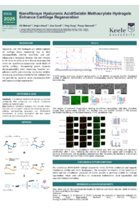
Introduction Various forms of hyaluronic acid (HA)-based hydrogels are widely utilized in cartilage tissue engineering due to their intrinsic bioactivity, biocompatibility, and anti-inflammatory properties. However, the high viscosity of HA solutions limits their processability into fibrous structures that mimic the nanofibrous nature of the hyaline cartilage extracellular matrix. To address this, gelatin is often incorporated to improve electrospinnability while simultaneously providing bioactive cell adhesion motifs and enhanced mechanical properties. In this study, a hyaluronic acid methacrylate/gelatin methacryloyl nanofibrous hydrogel was developed to mimic native ECM architecture and promote chondrogenic activity for cartilage regeneration.
Materials and Methods HA (200-500 kDa) and gelatin were methacrylated, achieving degrees of modification of approximately 18% and 60%, respectively, as confirmed by NMR. Lyophilized HAMA (1, 1.5 and 2% w/v) and GelMA (5, 7.5 and 10% w/v) were dissolved in hexafluoroisopropanol and assessed using the Box–Benkhen design of experiments model combined with rheological analysis to identify suitable concentrations and optimize electrospinning parameters. The optimized formulations were mixed with Irgacure 2959, electrospun into mats (~10 mm × 10 mm at ~2 mm thickness), and UV-crosslinked following immersion in pure ethanol. After crosslinking, the mats were rinsed with PBS and prepared for cell culture.
Results and Discussion SEM images showed the morphology of nanofibers, with diameters of approximately 300 nm for low-HAMA/high-GelMA formulations, whereas higher HAMA content resulted in thicker fibers in the 400–600 nm range. Confocal microscopy demonstrated the stability of mats for at least 21 days and high chondrocyte viability (>80%), indicating the absence of cytotoxic residues. Finally, GAG/DNA quantification and immunofluorescence staining confirmed the expression of type II collagen and aggrecan, resembling native hyaline cartilage.
Conclusion The results indicate that the HAMA/GelMA fibrous hydrogel represents a promising strategy for cartilage tissue engineering by combining the bioactivity of HAMA with the mechanical and cell-interactive properties of GelMA.
Keywords
Cartilage tissue engineering
Hyaluronic acid nanofibers
Hyaluronic acid electrospinning
nanofibrous hydrogels
Poster
Gels Poster.pdf
PREFORMULATORY STUDIES ON ROSMARINIC ACID NLCs FOR ORAL ADMINISTRATION
Gels based on natural polymers containing silver nanoprisms for combined chemo/phototherapy of cancer